. 2017 Sep;95(1-2):123-140.
doi: 10.1007/s11103-017-0641-5. Epub 2017 Jul 28.

A novel family of proline/serine-rich proteins, which are phospho-targets of stress-related mitogen-activated protein kinases, differentially regulates growth and pathogen defense in Arabidopsis thaliana

Affiliations

A novel family of proline/serine-rich proteins, which are phospho-targets of stress-related mitogen-activated protein kinases, differentially regulates growth and pathogen defense in Arabidopsis thaliana

Mieder Anthony Thomas Palm-Forster et al. Plant Mol Biol. 2017 Sep.

Abstract

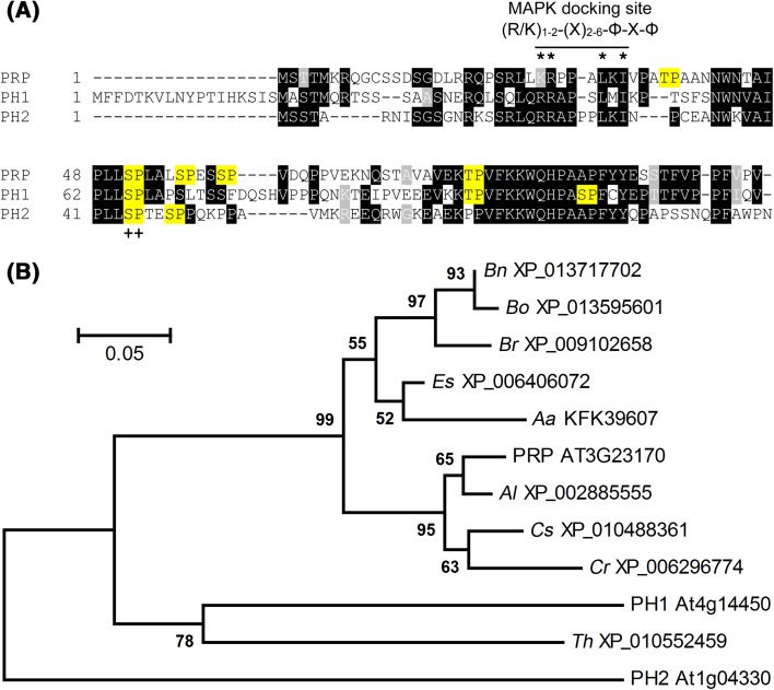
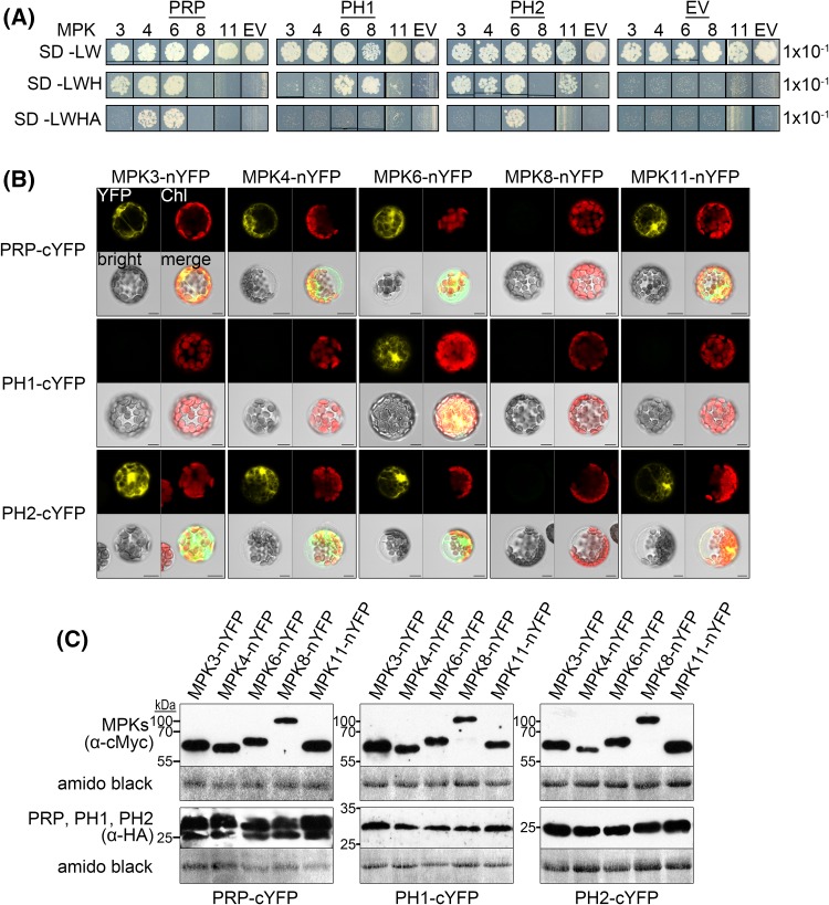
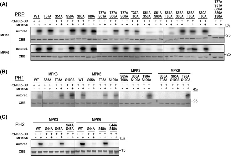
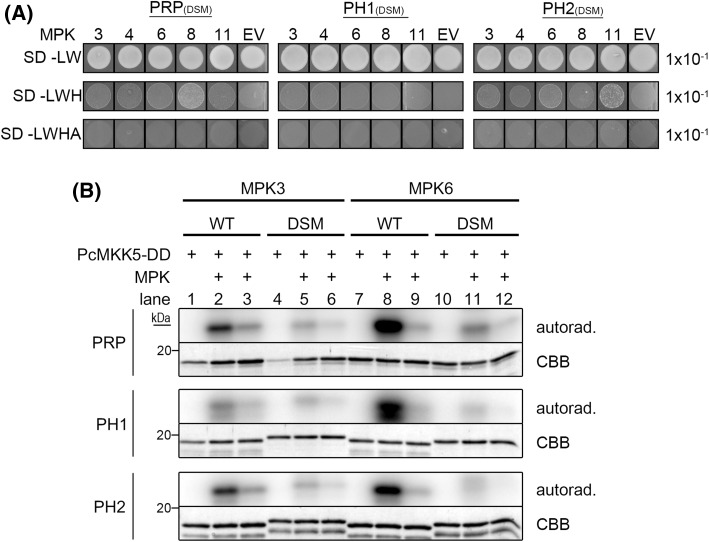
The molecular actions of mitogen-activated protein kinases (MAPKs) are ultimately accomplished by the substrate proteins where phosphorylation affects their molecular properties and function(s), but knowledge regarding plant MAPK substrates is currently still fragmentary. Here, we uncovered a previously uncharacterized protein family consisting of three proline/serine-rich proteins (PRPs) that are substrates of stress-related MAPKs. We demonstrated the importance of a MAPK docking domain necessary for protein-protein interaction with MAPKs and consequently also for phosphorylation. The main phosphorylated site was mapped to a residue conserved between all three proteins, which when mutated to a non-phosphorylatable form, differentially affected their protein stability. Together with their distinct gene expression patterns, this differential accumulation of the three proteins upon phosphorylation probably contributes to their distinct function(s). Transgenic over-expression of PRP, the founding member, led to plants with enhanced resistance to Pseudomonas syringae pv. tomato DC3000. Older plants of the over-expressing lines have curly leaves and were generally smaller in stature. This growth phenotype was lost in plants expressing the phosphosite variant, suggesting a phosphorylation-dependent effect. Thus, this novel family of PRPs may be involved in MAPK regulation of plant development and / or pathogen resistance responses. As datamining associates PRP expression profiles with hypoxia or oxidative stress and PRP-overexpressing plants have elevated levels of reactive oxygen species, PRP may connect MAPK and oxidative stress signaling.

Keywords: MAPK; Oxidative stress; PAMPs; Pathogen resistance; Phosphorylation; Plant development.


Figures

Fig. 1
Fig. 1
Sequence and phylogenetic analyses of PRP, PH1 and PH2. a Box shade representation of a multiple sequence alignment of PRP, PH1 and PH2. Putative MAPK phosphorylation sites ([S/T]P) are highlighted in yellow; ++ indicates a putative MAPK phosphorylation site conserved in all three proteins. Also indicated is a conserved MAPK docking site (R/K)1−2-(X)2−6-Φ-X-Φ, where Φ represents a hydrophobic residue, X stands for any residue (PRP: K26, R27, L31, I33; PH1: R42, R43, L47, I49; PH2: R21, R22, L27, I29). Molecular weights: PRP 11.7 kDa, PH1 14.1 kDa, PH2 11.3 kDa; proline content: PRP 15.9%, PH1 12.8%, PH2 18.0%. b Phylogenetic analysis of PRP, PH1, PH2 and close homologs from other species. GenBank identifiers are given next to the species abbreviations (Bn Brassica napus cv. ZS11, Bo Brassica oleracea var. oleracea, Br Brassica rapa cv. Chiifu-401-42, Es Eutrema salsugineum, Aa Arabis alpina cv. Pajares, Al Arabidopsis lyrata subsp. lyrata, Cs Camelina sativa cv. DH55, Cr Capsella rubella cv. Monte Gargano, Th Tarenaya hassleriana). Details are given in the “Materials and Methods”
Fig. 2
Fig. 2
PRP, PH1 and PH2 interact with MAPKs in vivo. a Yeast two-Hybrid (Y2H) interaction analyses of PRP, PH1 and PH2 proteins with Arabidopsis MAPKs. Positive interactions are indicated by yeast growth on selective synthetic drop-out media (SD-Leu/-Trp/-His or SD-Leu/-Trp/-His/-Ade); EV empty vector control (pDEST32). b Bimolecular fluorescence complementation (BiFC) assay in Arabidopsis protoplasts transiently expressing fusions of PRP, PH1 or PH2 with a C-terminal Yellow Fluorescent Protein (cYFP) fragment together with MPK3/4/6/8/11-nYFP. Positive interaction is indicated as fluorescence signal in the YFP channel; Chl chlorophyll autofluorescence, bright bright field, merge channel overlay. Scale bars 10 µm. c Western blot analyses of samples from b to monitor expression of the BiFC constructs. Samples were ran on duplicate gels for separate detection of MPKs (α-cMyc) and PRP/PH1/PH2 (α-HA). Membranes were stained with amido black to show equal loading (based on the staining of the Rubisco large subunit band). Experiments were performed three times with similar results
Fig. 3
Fig. 3
PRP, PH1 and PH2 are in vitro substrates of MPK3/6. a. Radioactive in vitro kinase assays with recombinant PRP wild type (WT) or putative MAPK phosphosite mutant proteins (individual, double and penta mutants) and MPK3/6. Activation of MPK3/6 was achieved in the presence of constitutively active parsley PcMKK5-DD (Lee et al. 2004). Proteins were incubated with radioactive ATP, separated by SDS-PAGE and analyzed by autoradiography (autorad.); CBB Coomassie Brilliant Blue stain (of the purified recombinant protein). Position of the protein size marker is indicated on the right. b and c Radioactive in vitro kinase assays with recombinant PH1/PH2 wild type (WT) or putative MAPK phosphosite mutant proteins (individual and double mutants) and MPK3/6. Samples were analyzed as in a. (This figure is previously part of Fig. 1d of the Palm-Forster et al. paper describing the mutagenesis method and is reprinted here with permission from Elsevier)
Fig. 4
Fig. 4
PRP, PH1 and PH2 have a functional MAPK docking site. a Yeast two-Hybrid (Y2H) interaction analyses of Arabidopsis MAPKs with PRP, PH1 and PH2 MAPK docking site mutant (DSM) versions (PRP: K26E, R27E, L31D, I33D; PH1: R42E, R43E, L47D, I49D; PH2: R21E, R22E, L27D, I29D). Positive interactions are indicated by yeast growth on selective synthetic drop-out media (SD-Leu/-Trp/-His or SD-Leu/-Trp/-His/-Ade; cf. Fig. 1a); EV empty vector control (pDEST32). b Radioactive in vitro kinase assays with recombinant PRP, PH1 and PH2 wild type (WT) or MAPK docking site mutant proteins (DSM) and either MPK3 or MPK6. Activation of MPK3/6 was achieved in the presence of constitutively active parsley PcMKK5-DD (Lee et al. 2004). Samples were analyzed as in Fig. 3a. For lanes 3, 6, 9 and 12, only 1/5 of MPK3/6 was used in comparison to lanes 2, 5, 8 and 11, respectively. Autorad. autoradiography, CBB Coomassie Brilliant Blue (staining of the recombinant substrate proteins)
Fig. 5
Fig. 5
PRP, PH1 and PH2 MAPK docking site mutants are impaired in MPK-interaction in vivo. a Bimolecular fluorescence complementation (BiFC) assay in Arabidopsis protoplasts transiently co-expressing cYFP fusions of PRP, PH1 or PH2 wild type (WT) or MAPK docking site mutants (DSM) together with MPK6-nYFP plus CFP-HA. Positive interaction is indicated as fluorescence signal in the YFP channel; CFP = CFP channel (for normalization of fluorescence levels); Chl chlorophyll autofluorescence; bright bright field. Scale bars 10 µm. As a measure for interaction, fluorescence ratios (YFP/CFP) were calculated for each individual protoplast (bar charts next to the microscopy images; n ≥ 35). b Western blot analyses of samples from a to monitor expression of the constructs. The blot was subsequently probed with α-cMyc (MPK6) and α-HA (PRP/PH1/PH2/CFP) antibodies. (Note that for unknown reasons, the DSM PH1 and PH2 show aberrant gel mobility compared to the non-mutated proteins; this is also seen with the recombinant proteins, see Fig. 4b). The membrane was stained with amido black to show equal loading. Experiments were repeated twice with similar results
Fig. 6
Fig. 6
PRP, PH1 and PH2 show a phospho-shift and/or destabilization upon phosphorylation. a–c PRP, PH1 and PH2 wild type, phosphosite mutant (alanine substitutions) or phospho-mimic (aspartic acid substitutions) proteins were transiently expressed in protoplasts. Upon elicitation with 100 nM flg22 alone or in combination with 1 µM cycloheximide (CHX), samples were harvested at the indicated time points and subjected to Western blot analyses using an α-HA antibody. Arrowheads indicate a phospho-shift of wild type PRP and PRP-S51D, as well as a double band of PH1-S65D. Note that PRP phosphosite mutants lost the phospho-shift upon flg22 treatment; mpt minutes post treatment. Membranes were stained with amido black to show equal loading. Position of a 25-kDa marker protein is indicated on the left of each blot
Fig. 7
Fig. 7
Expression and subcellular localization of PRP, PH1 and PH2. a Transient expression of PRP, PH1 and PH2 constructs tagged with N-terminal CFP or C-terminal GFP in mesophyll protoplasts (left panel) or Nicotiana benthamiana (right panel) for subcellular localization. Photos of CFP/GFP and chlorophyll autofluorescence (Chl) channels were taken. An empty vector construct (EV) served as a negative control. Scale bars 10 µm (left panel)/100 µM (right panel). b Expression analyses of PRP and its homologs in seedlings treated with the PAMPs, flg22 and elf18. Seedlings were grown in liquid culture for 2 weeks and treated with 1 µM flg22/elf18. Samples were harvested at the indicated time points. Total RNA was isolated, reverse transcribed and used for quantitative realtime PCR to measure PRP, PH1 and PH2 transcript levels (relative to PP2A as a reference gene). Asterisks indicate statistically significant differences compared to [t = 0 minutes] (n = 3; Kruskal–Wallis One-way ANOVA with Dunns posttest; *p < 0.05; **p < 0.01; ***p < 0.001). The experiment was repeated twice with similar results. c Luciferase assays in mesophyll protoplasts using PRP/PH1/PH2 promoter-luciferase fusions as reporter constructs. Luciferase-mediated light emission was recorded for 3 h post PAMP treatment (100 nM flg22/elf18) as a measure for promoter activity. For normalization, a pUBQ10-GUS construct was co-transformed. LUC/GUS ratios were calculated and fold changes relative to the respective water control [t = 0 minutes] were plotted. Letters indicate statistically significant differences (n = 3; Two-way RM ANOVA with Bonferroni posttests; p < 0.01; ns not significant). The experiment was performed four times with similar results
Fig. 8
Fig. 8
Characterization of transgenic plants over-expressing wild type or phosphosite mutant versions of PRP, PH1 and PH2. a Growth phenotype of plants grown for 8 weeks on soil under short day conditions (8 h light/16 h darkness, 22 °C). The right panel shows individual adult leaves. Two independent over-expressing lines per genotype are shown (OE1/OE2). Note that only the main individual phosphosites were mutated in the phosphosite mutants (PRP-S51A, PH1-S65A, PH2-S44A; cf. Fig. 2). b Western blot (α-cMyc) showing expression of the constructs in the transgenic lines in a. Membranes were stained with amido black to show equal loading (WT wild type, Pmut phosphosite mutants). c Root growth inhibition assays with seedlings grown on plates ±1 µM flg22. The experiment was performed twice with similar results and the combined data are shown. Asterisks indicate statistically significant differences (Mann–Whitney test; **p < 0.01; ***p < 0.001). d Infection assay with Pseudomonas syringae pv. tomato (Pst) DC3000. Plants were grown for 3 weeks on soil under short day conditions (8 h light/16 h darkness) and then spray-inoculated with a bacterial solution (5 × 108 cells/ml). Bacterial growth was determined (0 and 3 dpi) by counting colony-forming units (CFU) after plating serial dilutions. The experiment was performed 5 times with similar results (the diagram shows combined data sets; n = 15). Asterisks indicate statistically significant differences at day 3 between over-expressing lines and the wild type (Col-0; Mann–Whitney test; *p < 0.05; ***p < 0.001; ns not significant). On the left side, photos of infected plants at day 3 are shown
Fig. 9
Fig. 9
ROS accumulation and PAD3 expression are enhanced in the PRP-overexpressors. a Flg22-induced H2O2 accumulation was quantified using a luminol-based assay. Four independent experiments were performed with different batches of plants, each with 24 leaf discs (Black = Col-0; red/blue = PRP overexpressor line 1 and 2, respectively). For the depicted graph, data were pooled from all four experiments (n = 4 × 24 leaf discs) and reported as the mean relative light units (RLU). Error bars are the standard errors. Inset shows the basal ROS levels prior to flg22 treatment. Different alphabets mark statistically distinct (ANOVA) groups. b qRT-PCR showing expression of the indicated defense-related genes. Flg22 (100 nM), H2O2 (2 mM) or water (mock) treatments were performed for 30 min and the RNA extracted for RT-PCR analysis. Data is pooled (n = 9) from three independently performed experiments (each with 3 replicates) and shown as relative expression levels (normalized to the average of the Col-0/mock-treated sample). For statistical significance test, the data was log2-transformed and the p-values of the pair-wise t test comparison (to the corresponding Col-0 genotype) are shown above each bar. c Expression of PRP, PH1 and PH2 was analyzed as in “b” above

Similar articles

Cited by

References

    1. Andreasson E, et al. The MAP kinase substrate MKS1 is a regulator of plant defense responses. EMBO J. 2005;24:2579–2589. doi: 10.1038/sj.emboj.7600737. - DOI - PMC - PubMed
    1. Asai T, et al. MAP kinase signalling cascade in Arabidopsis innate immunity. Nature. 2002;415:977–983. doi: 10.1038/415977a. - DOI - PubMed
    1. Benschop JJ, Mohammed S, O’, Flaherty M, Heck AJ, Slijper M, Menke FL. Quantitative phosphoproteomics of early elicitor signaling in Arabidopsis. Mol Cell Proteomics. 2007;6:1198–1214. doi: 10.1074/mcp.M600429-MCP200. - DOI - PubMed
    1. Bethke G, Unthan T, Uhrig JF, Pöschl Y, Gust AA, Scheel D, Lee J. Flg22 regulates the release of an ethylene response factor substrate from MAP kinase 6 in Arabidopsis thaliana via ethylene signaling. Proc Natl Acad Sci U S A. 2009;106:8067–8072. doi: 10.1073/pnas.0810206106. - DOI - PMC - PubMed
    1. Bethke G, et al. Activation of the Arabidopsis thaliana mitogen-activated protein kinase MPK11 by the flagellin-derived elicitor peptide, flg22. Mol Plant Microbe Interact. 2012;25:471–480. doi: 10.1094/MPMI-11-11-0281. - DOI - PubMed

MeSH terms