

Targeted dianthin is a powerful toxin to treat pancreatic carcinoma when applied in combination with the glycosylated triterpene SO1861
- PMID: 28755527
- PMCID: PMC5664001
- DOI: 10.1002/1878-0261.12115
Targeted dianthin is a powerful toxin to treat pancreatic carcinoma when applied in combination with the glycosylated triterpene SO1861
Abstract
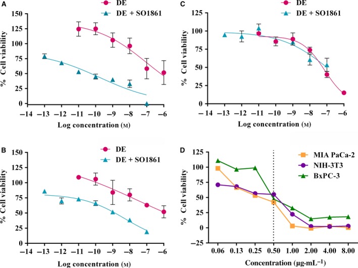
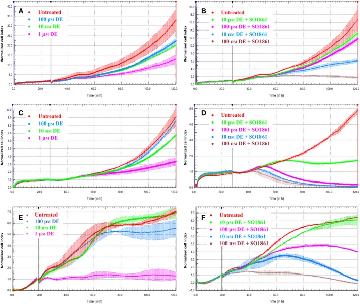
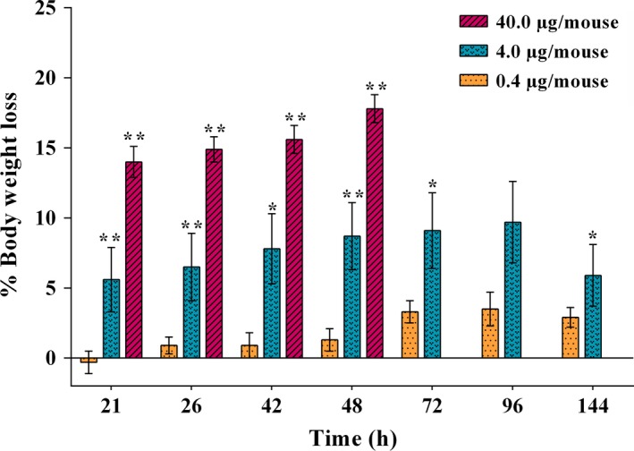
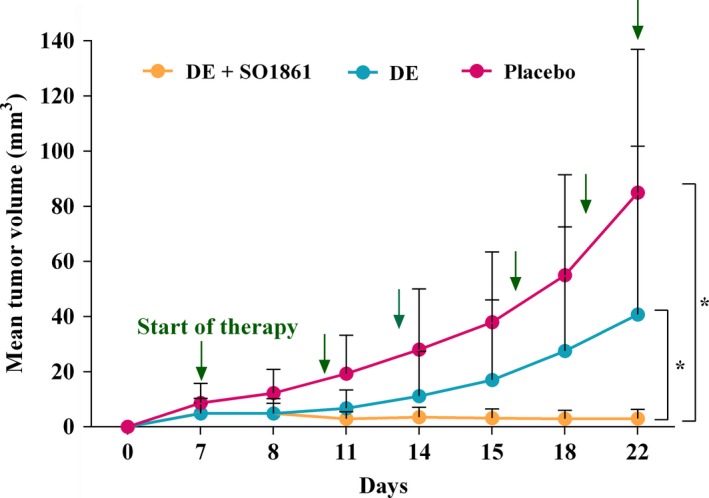
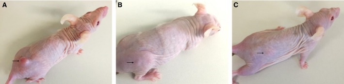
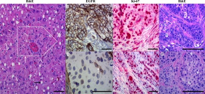
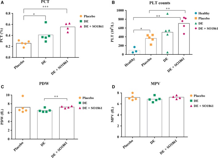
Targeted cancer therapy provides the basis for the arrest of tumor growth in aggressive pancreatic carcinoma; however, a number of protein-based targeted toxins lack efficacy due to insufficient endosomal escape after being endocytosed. Therefore, we tested a fusion protein of the ribosome-inactivating protein dianthin and human epidermal growth factor in combination with a glycosylated triterpene (SO1861) that serves as an endosomal escape enhancer. In vitro investigations with the pancreatic carcinoma cell lines BxPC-3 and MIA PaCa-2 revealed no significant differences to off-target cells in the half maximal inhibitory concentration (IC50 ) for the fusion protein. In contrast, combination with SO1861 decreased the IC50 for BxPC-3 cells from 100 to 0.17 nm, whereas control cells remained unaffected. Monotherapy of BxPC-3 xenografts in CD-1 nude mice led to a 51.7% average reduction in tumor size (40.8 mm3 ) when compared to placebo; however, combined treatment with SO1861 resulted in a more than 13-fold better efficacy (3.0 mm3 average tumor size) with complete regression in 80% of cases. Immunohistochemical analyses showed that tumor cells with lower target receptor expression are, in contrast to the combination therapy, able to escape from the monotherapy, which finally results in tumor growth. At the effective concentration, we did not observe liver toxicity and saw no other side effects with the exception of a reversible skin hardening at the SO1861 injection site, alongside an increase in platelet counts, plateletcrit, and platelet distribution width. In conclusion, combining a targeted toxin with SO1861 is proven to be a very promising approach for pancreatic cancer treatment.
Keywords: SO1861; endosomal escape; epidermal growth factor receptor; pancreatic carcinoma; targeted toxin; xenograft.
© 2017 The Authors. Published by FEBS Press and John Wiley & Sons Ltd.
Figures
Similar articles
-
Improved Therapy of B-Cell Non-Hodgkin Lymphoma by Obinutuzumab-Dianthin Conjugates in Combination with the Endosomal Escape Enhancer SO1861.Toxins (Basel). 2022 Jul 13;14(7):478. doi: 10.3390/toxins14070478. Toxins (Basel). 2022. PMID: 35878216 Free PMC article.
-
Combinatorial approach to increase efficacy of Cetuximab, Panitumumab and Trastuzumab by dianthin conjugation and co-application of SO1861.Biochem Pharmacol. 2015 Oct 1;97(3):247-55. doi: 10.1016/j.bcp.2015.07.040. Epub 2015 Aug 5. Biochem Pharmacol. 2015. PMID: 26253687
-
Magnetic Nanoparticle-Based Dianthin Targeting for Controlled Drug Release Using the Endosomal Escape Enhancer SO1861.Nanomaterials (Basel). 2021 Apr 20;11(4):1057. doi: 10.3390/nano11041057. Nanomaterials (Basel). 2021. PMID: 33924180 Free PMC article.
-
Dianthin and Its Potential in Targeted Tumor Therapies.Toxins (Basel). 2019 Oct 11;11(10):592. doi: 10.3390/toxins11100592. Toxins (Basel). 2019. PMID: 31614697 Free PMC article. Review.
-
Saponins as tool for improved targeted tumor therapies.Curr Drug Targets. 2009 Feb;10(2):140-51. doi: 10.2174/138945009787354584. Curr Drug Targets. 2009. PMID: 19199910 Review.
Cited by
-
Saponin Fraction CIL1 from Lysimachia ciliata L. Enhances the Effect of a Targeted Toxin on Cancer Cells.Pharmaceutics. 2023 Apr 28;15(5):1350. doi: 10.3390/pharmaceutics15051350. Pharmaceutics. 2023. PMID: 37242592 Free PMC article.
-
Mutational Analysis of RIP Type I Dianthin-30 Suggests a Role for Arg24 in Endocytosis.Toxins (Basel). 2024 May 10;16(5):219. doi: 10.3390/toxins16050219. Toxins (Basel). 2024. PMID: 38787071 Free PMC article.
-
Improved Therapy of B-Cell Non-Hodgkin Lymphoma by Obinutuzumab-Dianthin Conjugates in Combination with the Endosomal Escape Enhancer SO1861.Toxins (Basel). 2022 Jul 13;14(7):478. doi: 10.3390/toxins14070478. Toxins (Basel). 2022. PMID: 35878216 Free PMC article.
-
A cleavable peptide adapter augments the activity of targeted toxins in combination with the glycosidic endosomal escape enhancer SO1861.BMC Biotechnol. 2024 Apr 29;24(1):24. doi: 10.1186/s12896-024-00854-5. BMC Biotechnol. 2024. PMID: 38685061 Free PMC article.
-
Glycosylated Triterpenoids as Endosomal Escape Enhancers in Targeted Tumor Therapies.Biomedicines. 2017 Mar 29;5(2):14. doi: 10.3390/biomedicines5020014. Biomedicines. 2017. PMID: 28536357 Free PMC article. Review.
References
-
- Ali S, El‐Rayes BF, Sarkar FH and Philip PA (2005) Simultaneous targeting of the epidermal growth factor receptor and cyclooxygenase‐2 pathways for pancreatic cancer therapy. Mol Cancer Ther 4, 1943–1951. - PubMed
-
- Bachran C, Durkop H, Sutherland M, Bachran D, Muller C, Weng A, Melzig MF and Fuchs H (2009) Inhibition of tumor growth by targeted toxins in mice is dramatically improved by saponinum album in a synergistic way. J Immunother 32, 713–725. - PubMed
-
- Bachran D, Schneider S, Bachran C, Urban R, Weng A, Melzig MF, Hoffmann C, Kaufmann AM and Fuchs H (2010b) Epidermal growth factor receptor expression affects the efficacy of the combined application of saponin and a targeted toxin on human cervical carcinoma cells. Int J Cancer 127, 1453–1461. - PubMed
-
- Bachran D, Schneider S, Bachran C, Weng A, Melzig MF and Fuchs H (2011) The endocytic uptake pathways of targeted toxins are influenced by synergistically acting Gypsophila saponins. Mol Pharm 8, 2262–2272. - PubMed
Publication types
MeSH terms
Substances
LinkOut - more resources
Full Text Sources
Other Literature Sources
Medical
Research Materials
