. 2018 Feb;14(2):243-252.
doi: 10.1016/j.jalz.2017.04.015. Epub 2017 Jul 26.

Peripheral complement interactions with amyloid β peptide in Alzheimer's disease: 2. Relationship to amyloid β immunotherapy

Affiliations

Peripheral complement interactions with amyloid β peptide in Alzheimer's disease: 2. Relationship to amyloid β immunotherapy

Andrés Crane et al. Alzheimers Dement. 2018 Feb.

Abstract

Introduction: Our previous studies have shown that amyloid β peptide (Aβ) is subject to complement-mediated clearance from the peripheral circulation, and that this mechanism is deficient in Alzheimer's disease. The mechanism should be enhanced by Aβ antibodies that form immune complexes (ICs) with Aβ, and therefore may be relevant to current Aβ immunotherapy approaches.

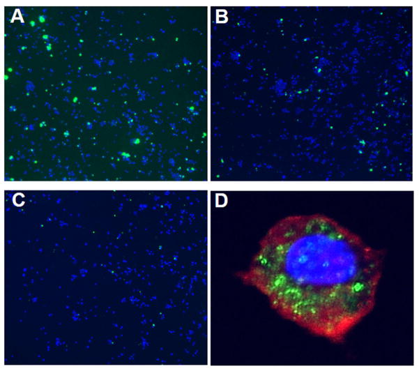
Methods: Multidisciplinary methods were employed to demonstrate enhanced complement-mediated capture of Aβ antibody immune complexes compared with Aβ alone in both erythrocytes and THP1-derived macrophages.

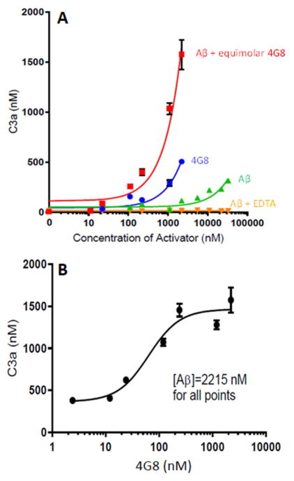
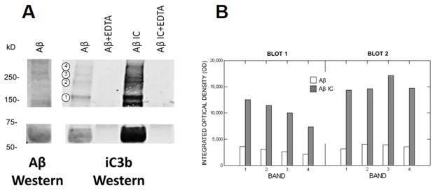
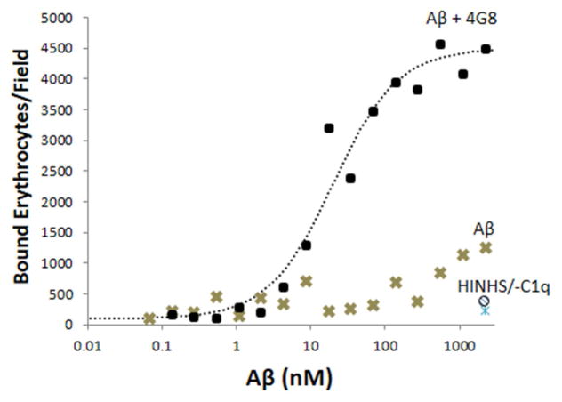
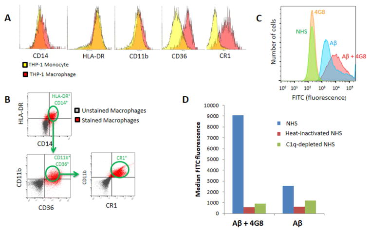
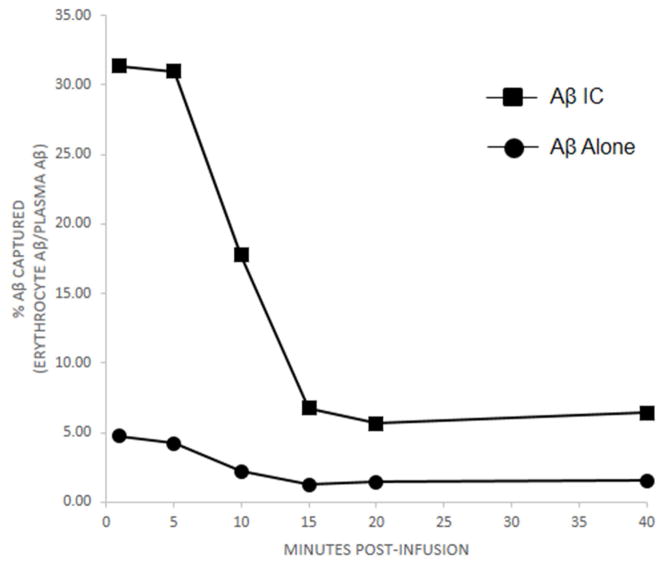
Results: Aβ antibodies dramatically increased complement activation and opsonization of Aβ, followed by commensurately enhanced Aβ capture by human erythrocytes and macrophages. These in vitro findings were consistent with enhanced peripheral clearance of intravenously administered Aβ antibody immune complexes in nonhuman primates.

Discussion: Together with our previous results, showing significant Alzheimer's disease deficits in peripheral Aβ clearance, the present findings strongly suggest that peripheral mechanisms should not be ignored as contributors to the effects of Aβ immunotherapy.

Keywords: Alzheimer's disease; Amyloid β peptide; Aβ immunotherapy; Blood; Complement; Complement receptor 1; Erythrocyte; Human; Macrophage.


Conflict of interest statement

CONFLICTS OF INTEREST: None

Figures

Fig. 1
Fig. 1. Complement activation by Aβ and Aβ/4G8 ICs
A) Aβ42 was incubated with equimolar Aβ antibody 4G8 to form Aβ/4G8 ICs, then reacted with NHS as a complement source. Significant dose-dependent complement activation (P < 0.001), as measured by C3a generated, was observed for Aβ ICs even at low nM concentrations. Aβ alone and 4G8 alone also dose-dependently activated complement (P < 0.001), but required μM concentrations to stimulate C3a production above background levels. EDTA abolished these effects, showing that they are specific to complement mechanisms. B) Complement activation by Aβ ICs could also be shown to be dose-dependent with respect to the amount of Aβ antibody available (P < 0.001). Here, a constant amount of Aβ (2215 nM) was incubated with varying concentrations of Aβ antibody 4G8, from low to equimolar concentrations relative to Aβ. Note that the X-axis is log-scaled in these graphs to include the wide range of concentrations. For both figures, each data point represents the mean of triplicate samples. Error bars denote standard error of the mean. Standard error for replicates is not shown when they are smaller than the symbols for a particular concentration.
Fig. 2
Fig. 2. Complement opsonization of Aβ and Aβ/4G8 ICs
A) After exposure to NHS to permit complement activation, Aβ alone and Aβ IC solutions were run on SDS reducing/denaturing Western blots to detect the complement opsonin iC3b. EDTA treatment (lanes 2 and 4 of the iC3b blot) abolished complement activation. The absence of iC3b immunoreactivity after EDTA treatment therefore shows that the iC3b detected in Aβ alone and Aβ IC conditions (lanes 1 and 3 of the iC3b blot) was specifically generated by Aβ and Aβ IC complement activation, and not by endogenous levels of iC3b. Compared to Aβ alone, iC3b immunoreactivity was markedly enhanced by Aβ ICs, consistent with enhanced complement activation by Aβ ICs in our earlier experiments. A parallel Western blot for Aβ (6E10 detection antibody) using the Aβ solution employed in the experiment is shown in the left-most lane. Note that bands for iC3b and Aβ aggregate forms typically coincide, as would be predicted given that iC3b is covalently bound to Aβ aggregate forms that can activate complement. B) Densitometry of the blot in panel A, as well as a second blot, prepared in the same way but pre-depleted of endogenous Igs and iC3b, gave a similar pattern of results that did not differ significantly from blot to blot (P = 0.307), with both showing highly-enriched iC3b immunoreactivity in Aβ IC compared to Aβ alone samples (F = 71.65, P = 0.001). For example, integrated optical density of the molecular weight bands designated by the four circled numbers on panel A were increased by 3-fold to 5-fold in the Aβ IC condition.
Fig. 3
Fig. 3. Erythrocyte capture of Aβ and Aβ ICs
In erythrocyte adhesion assays, erythrocytes bound to Aβ-coated and Aβ/4G8 IC-coated plates in a dose-dependent manner (P < 0.001) and binding was significantly greater for Aβ ICs (P < 0.001). Abolition of effects by heat-inactivated NHS at the highest dose of Aβ ICs and Aβ alone (HINHS and ○ symbol at bottom right of graph) demonstrated specificity to complement reactions as opposed to erythrocyte binding by mechanisms other than complement. Abolition of effects by C1q-depleted serum (-C1q and △ symbol at bottom right of graph) confirmed this finding and, in addition, strongly suggested that erythrocyte capture of Aβ is primarily mediated by the classical, rather than the alternative complement pathway. Each data point represents the mean of duplicates. The vast majority of data points exhibited standard errors that were too small to show clearly. For the Aβ alone condition, the average standard error of the mean was 111.3 bound erythrocytes/field (range of 7.8 to 422.5). For the Aβ IC condition, the average standard error of the mean was 321.0 bound erythrocytes/field (range of 9.9 to 996.7).
Fig. 4
Fig. 4. Macrophage capture of Aβ and Aβ ICs
A) Flow cytometry characterization of THP-1 monocytes and their differentiation to THP-1-derived macrophages revealed a transition to increased expression of inflammatory markers such as CD36, a mediator of macrophage phagocytosis [25], and, especially, CR1, the macrophage receptor for the major complement opsonins in primates [34,35]. B) To insure assessment of the macrophage-specific phenotype in subsequent experiments, all cells were gated for criterion expression of five different macrophage markers, CD14, HLA-DR, CD11B, CD36, and CR1. C) Fluorescence intensity distributions for samples pre-incubated with NHS. Distributions designated NHS and 4G8 represent fluorescence of the macrophage markers on which the cells were gated. Fluorescence distributions for gated, FITC-labeled Aβ and Aβ IC samples appear in the right-hand portion of the graph. A total of 100,000 cells were evaluated for each sample. D) Median fluorescence intensity of Aβ captured by THP-1-derived macrophages increased nearly 4-fold for Aβ ICs compared to Aβ alone. Only background readings were obtained for the cells when heat-inactivated NHS or C1q-depleted serum were substituted for NHS as a complement source.
Fig. 5
Fig. 5. Macrophage uptake of Aβ and Aβ ICs
A–C) Representative fluorescence micrographs (10X) of THP-1-derived macrophages (DAPI nuclear counterstain) (blue) after exposure to 1 μM FITC-conjugated Aβ plus 0.1 μM Aβ antibody 4G8 to form Aβ ICs (A) or 1 μM FITC-conjugated Aβ alone (B). Aβ IC and Aβ alone solutions were previously incubated with NHS to permit complement activation. The number of cells co-localized with Aβ (green), as well as their fluorescence intensity, was clearly enhanced by Aβ ICs. Heat inactivation of the serum reduced staining to background (C). D) Representative high power (40X objective) confocal micrograph of a macrophage after exposure to FITC-conjugated Aβ plus 4G8 and NHS. PKH26 (red) was used to stain the cell membrane and DAPI (blue) was used to stain the nucleus. Localization of the FITC label for Aβ (green) was primarily intracellular, suggesting that the macrophages had phagocytosed the Aβ ICs.
Fig. 6
Fig. 6. Erythrocyte uptake and clearance of Aβ and Aβ ICs in non-human primates
Relative to the amount of Aβ available in the plasma compartment, erythrocyte uptake and clearance of Aβ inoculated into two non-human primates was significantly greater for Aβ ICs compared to Aβ alone (P < 0.001). Because erythrocyte and plasma levels at baseline differed in the animals, the data in the figure have been normalized as percent of Aβ recovered in the erythrocyte compartment relative to the amount available in the plasma compartment. Clearance into the erythrocyte compartment was rapid over the first 15 minutes after Aβ or Aβ IC injection, a characteristic of immune adherence [41]. By 20 minutes, Aβ IC values had returned to baseline, but remained elevated by some 20% relative to baseline for Aβ alone. Samples at each time point in each animal were assayed in duplicate. Standard error bars for each time point for each animal were smaller than the symbols at each time point and are not shown. For Aβ alone, mean standard error was 0.13% (range of 0.01 to 0.39). For Aβ IC, mean standard error was 0.82% (range of 0.10 to 2.81).

Similar articles

Cited by

References

    1. Lannfelt L, Relkin NR, Siemers ER. Amyloid-β-directed immunotherapy for Alzheimer’s disease. J Intern Med. 2014 Mar;275(3):284–95. - PMC - PubMed
    1. Bussière T, Bard F, Barbour R, Grajeda H, Guido T, et al. Morphological characterization of Thioflavin-S-positive amyloid plaques in transgenic Alzheimer mice and effect of passive Abeta immunotherapy on their clearance. Am J Pathol. 2004 Sep;165(3):987–95. - PMC - PubMed
    1. Bard F, Cannon C, Barbour R, Burke RL, Games D, et al. Peripherally administered antibodies against amyloid beta-peptide enter the central nervous system and reduce pathology in a mouse model of Alzheimer disease. Nat Med. 2000 Aug;6(8):916–9. - PubMed
    1. Gilman S, Koller M, Black RS, et al. Clinical effects of A-beta immunization (AN1792) in patients with AD in an interrupted trial. Neurology. 2005;64(9):1553–1562. - PubMed
    1. Boche D, Denham N, Holmes C, Nicoll JA. Neuropathology after active Abeta42 immunotherapy: implications for Alzheimer’s disease pathogenesis. Acta Neuropathol. 2010 Sep;120(3):369–84. - PubMed

Publication types

MeSH terms