A case of deep infection after instrumentation in dorsal spinal surgery: the management with antibiotics and negative wound pressure without removal of fixation
- PMID: 28756380
- PMCID: PMC5623226
- DOI: 10.1136/bcr-2017-220792
A case of deep infection after instrumentation in dorsal spinal surgery: the management with antibiotics and negative wound pressure without removal of fixation
Abstract
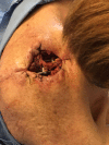
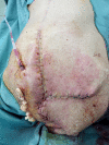
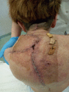
Until today the role of spinal instrumentation in the presence of a wound infection has been widely discussed and recently many authors leave the hardware in place with appropriate antibiotic therapy. This is a case of a 65-year-old woman suffering from degenerative scoliosis and osteoporotic multiple vertebral collapses treated with posterior dorsolumbar stabilisation with screws and rods. Four months later, skin necrosis and infection appeared in the cranial wound with exposure of the rods. A surgical procedure of debridement of the infected tissue and package with a myocutaneous trapezius muscle flap was performed. One week after surgery, negative pressure wound therapy was started on the residual skin defect. The wound healed after 2 months. The aim of this case report is to focus on the utility of this method even in the case of hardware exposure and infection. This may help avoid removing instrumentation and creating instability.
Keywords: Infection (neurology); Infections; Spinal Cord; Surgery.
© BMJ Publishing Group Ltd (unless otherwise stated in the text of the article) 2017. All rights reserved. No commercial use is permitted unless otherwise expressly granted.
Conflict of interest statement
Competing interests: None declared.
Figures


Similar articles
-
Epidural abscess as a delayed complication of spinal instrumentation in scoliosis surgery: a case of progressive neurologic dysfunction with complete recovery.Spine (Phila Pa 1976). 2008 Feb 1;33(3):E76-80. doi: 10.1097/BRS.0b013e31816245a6. Spine (Phila Pa 1976). 2008. PMID: 18303449
-
Treatment of postoperative infection after posterior spinal fusion and instrumentation in a patient with neuromuscular scoliosis.Am J Orthop (Belle Mead NJ). 2014 Feb;43(2):89-93. Am J Orthop (Belle Mead NJ). 2014. PMID: 24551867 Review.
-
Vacuum-assisted closure for deep infection after spinal instrumentation for scoliosis.J Bone Joint Surg Br. 2008 Mar;90(3):377-81. doi: 10.1302/0301-620X.90B3.19890. J Bone Joint Surg Br. 2008. PMID: 18310764
-
Delayed infection after elective spinal instrumentation and fusion. A retrospective analysis of eight cases.Spine (Phila Pa 1976). 1997 Oct 15;22(20):2444-50; discussion 2450-1. doi: 10.1097/00007632-199710150-00023. Spine (Phila Pa 1976). 1997. PMID: 9355228
-
Management of spinal infections in children with cerebral palsy.Orthop Traumatol Surg Res. 2016 Oct;102(6):801-5. doi: 10.1016/j.otsr.2016.04.015. Epub 2016 Jul 29. Orthop Traumatol Surg Res. 2016. PMID: 27480292 Review.
Cited by
-
Effectiveness of negative pressure wound therapy in treating deep surgical site infections after spine surgery: a meta-analysis of single-arm studies.J Orthop Surg Res. 2025 Jan 13;20(1):44. doi: 10.1186/s13018-025-05463-2. J Orthop Surg Res. 2025. PMID: 39800681 Free PMC article.
-
Ultra-early surgery in complete cervical spinal cord injury improves neurological recovery: A single-center retrospective study.Surg Neurol Int. 2019 Oct 18;10:207. doi: 10.25259/SNI_485_2019. eCollection 2019. Surg Neurol Int. 2019. PMID: 31768287 Free PMC article.
-
The relationship between preoperative predictive factors for clinical outcome in patients operated for lumbar spinal stenosis by decompressive laminectomy.Surg Neurol Int. 2020 Feb 25;11:27. doi: 10.25259/SNI_583_2019. eCollection 2020. Surg Neurol Int. 2020. PMID: 32123615 Free PMC article.
-
Rare case of anterior cervical discectomy and fusion complication in a patient with Zenker's diverticulum.BMJ Case Rep. 2018 Dec 9;11(1):e226022. doi: 10.1136/bcr-2018-226022. BMJ Case Rep. 2018. PMID: 30567215 Free PMC article.
-
Evaluation of prognostic preoperative factors in patients undergoing surgery for spinal metastases: Results in a consecutive series of 81 cases.Surg Neurol Int. 2022 Aug 19;13:363. doi: 10.25259/SNI_276_2022. eCollection 2022. Surg Neurol Int. 2022. PMID: 36128147 Free PMC article.
References
Publication types
MeSH terms
Substances
LinkOut - more resources
Full Text Sources
Other Literature Sources
Medical
