

EPR Oximetry of Cetuximab-Treated Head-and-Neck Tumours in a Mouse Model
- PMID: 28756482
- PMCID: PMC5691101
- DOI: 10.1007/s12013-017-0814-5
EPR Oximetry of Cetuximab-Treated Head-and-Neck Tumours in a Mouse Model
Abstract
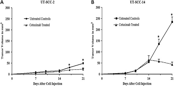
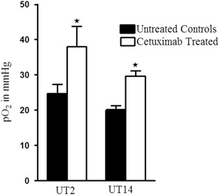
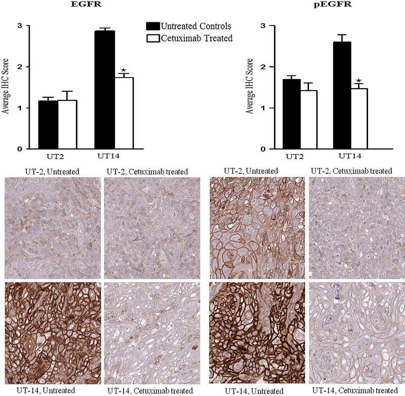
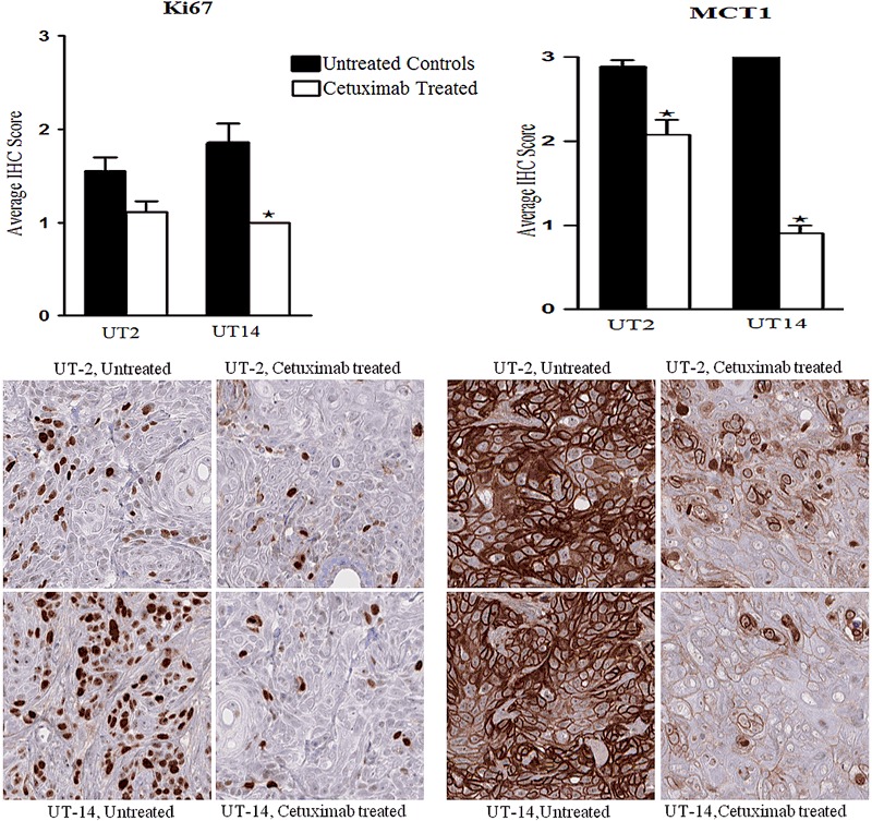
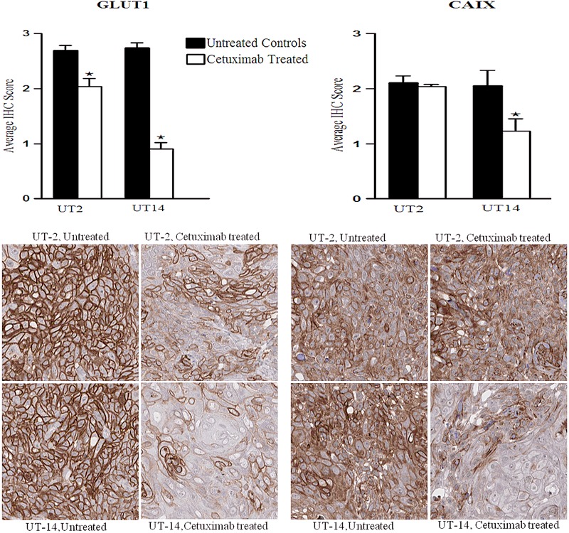
Head and neck squamous cell carcinoma (HNSCC) tumours are associated with high mortality despite advances in therapy. The monoclonal antibody cetuximab (Erbitux®) has been approved for the treatment of advanced HNSCC. However, only a subset of HNSC patients receiving cetuximab actually responds to treatment, underlining the need for a means to tailor treatments of individual patients. The aim of the present study was to investigate the effect of cetuximab treatment on tumour growth, on tumour partial oxygen pressure as measured by LiPc electron paramagnetic resonance oximetry and on the expression of proteins involved in tumour growth, metabolism and hypoxia. Two HNSCC cell lines, UT-SCC-2 and UT-SCC-14, were used to generate xenografts on female BALB/c (nu/nu) nude mice. Mice with xenografts were given three injections of intraperitoneal cetuximab or phosphate-buffered saline, and the tumour volume was recorded continuously. After treatment the tumour partial oxygen pressure was measured by LiPc electron paramagnetic resonance oximetry and the expression of epidermal growth factor receptor (EGFR), phosphorylated EGFR, Ki-67, MCT1, MCT4, GLUT1, CAIX and HIF-1α were investigated by immunohistochemistry. In xenografts from both cell lines (UT-SCC-2 and UT-SCC-14) cetuximab had effect on the tumour volume but the effect was more pronounced on UT-SCC-14 xenografts. A higher tumour oxygenation was measured in cetuximab-treated tumours from both cell lines compared to untreated controls. Immunocytochemical staining after cetuximab treatment shows a significantly decreased expression of EGFR, pEGFR, Ki67, CAIX and nuclear HIF-1α in UT-SCC-14 tumours compared to untreated controls. MCT1 and GLUT1 were significantly decreased in tumours from both cell lines but more pronounced in UT-SCC-14 tumours. Taken together, our results show that cetuximab treatment decreases the tumour growth and increases the tumour partial oxygen pressure of HNSCC xenografts. Furthermore we found a potential connection between the partial oxygen pressure of the tumours and the expression of proteins involved in tumour growth, metabolism and hypoxia.
Keywords: Cetuximab; EPR oximetry; Head and neck cancer; Hypoxia; Metabolism; Tumour oxygen pressure.
Conflict of interest statement
The authors declare that they have no competing interests.
Figures

Similar articles
-
Cetuximab sensitivity of head and neck squamous cell carcinoma xenografts is associated with treatment-induced reduction in EGFR, pEGFR, and pSrc.J Oral Pathol Med. 2017 Oct;46(9):717-724. doi: 10.1111/jop.12545. Epub 2017 Jan 28. J Oral Pathol Med. 2017. PMID: 28036101
-
Hypoxia Mediates Differential Response to Anti-EGFR Therapy in HNSCC Cells.Int J Mol Sci. 2017 Apr 29;18(5):943. doi: 10.3390/ijms18050943. Int J Mol Sci. 2017. PMID: 28468237 Free PMC article.
-
Epidermal growth factor receptor inhibition reduces angiogenesis via hypoxia-inducible factor-1α and Notch1 in head neck squamous cell carcinoma.PLoS One. 2015 Feb 27;10(2):e0119723. doi: 10.1371/journal.pone.0119723. eCollection 2015. PLoS One. 2015. PMID: 25723392 Free PMC article.
-
EGFR targeting drugs in the treatment of head and neck squamous cell carcinoma.Expert Opin Emerg Drugs. 2010 Jun;15(2):185-201. doi: 10.1517/14728211003716442. Expert Opin Emerg Drugs. 2010. PMID: 20415599 Review.
-
Advanced Squamous Cell Carcinoma of the Head and Neck: The Current Role of Cetuximab.ORL J Otorhinolaryngol Relat Spec. 2016;78(6):320-333. doi: 10.1159/000455891. Epub 2017 Jan 27. ORL J Otorhinolaryngol Relat Spec. 2016. PMID: 28125819 Review.
Cited by
-
The Role of Glucose Transporters in Oral Squamous Cell Carcinoma.Biomolecules. 2021 Jul 21;11(8):1070. doi: 10.3390/biom11081070. Biomolecules. 2021. PMID: 34439735 Free PMC article.
-
Comparison of phosphorescent agents for noninvasive sensing of tumor oxygenation via Cherenkov-excited luminescence imaging.J Biomed Opt. 2019 Mar;24(3):1-8. doi: 10.1117/1.JBO.24.3.036001. J Biomed Opt. 2019. PMID: 30834723 Free PMC article.
References
-
- Ang KK, Berkey BA, Tu X, et al. Impact of epidermal growth factor receptor expression on survival and pattern of relapse in patients with advanced head and neck carcinoma. Cancer Research. 2002;62:7350–7356. - PubMed
MeSH terms
Substances
LinkOut - more resources
Full Text Sources
Other Literature Sources
Medical
Research Materials
Miscellaneous
