. 2017 Aug 7;27(15):2365-2373.e8.
doi: 10.1016/j.cub.2017.06.069. Epub 2017 Jul 27.

Expanded Satellite Repeats Amplify a Discrete CENP-A Nucleosome Assembly Site on Chromosomes that Drive in Female Meiosis

Affiliations

Expanded Satellite Repeats Amplify a Discrete CENP-A Nucleosome Assembly Site on Chromosomes that Drive in Female Meiosis

Aiko Iwata-Otsubo et al. Curr Biol. .

Abstract

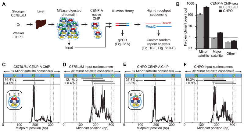
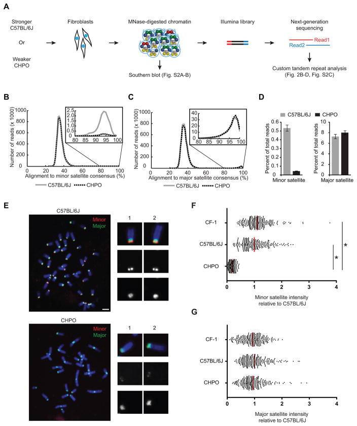
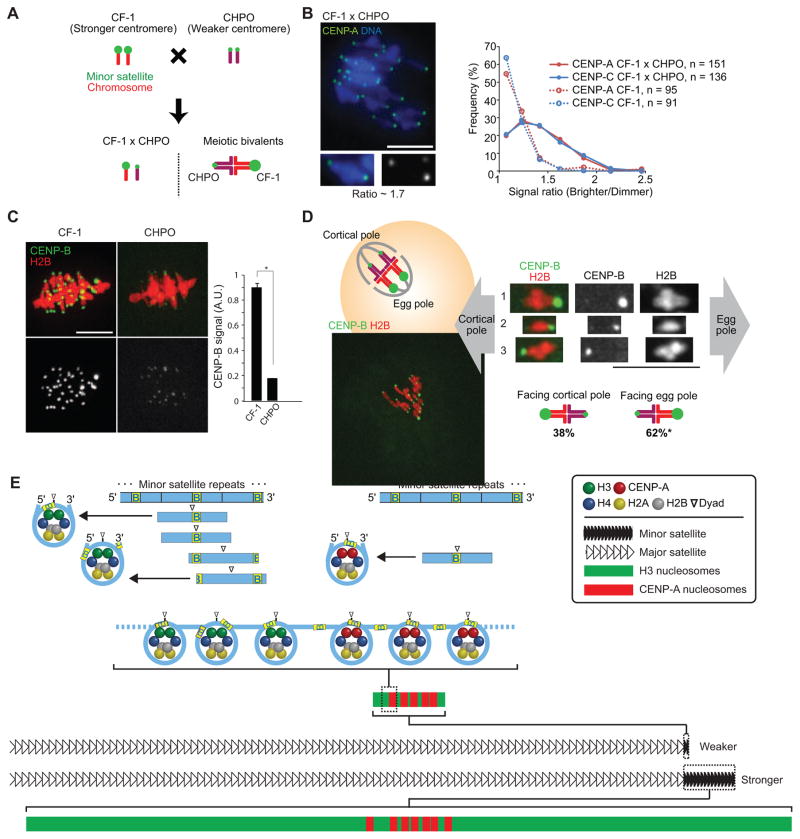
Female meiosis provides an opportunity for selfish genetic elements to violate Mendel's law of segregation by increasing the chance of segregating to the egg [1]. Centromeres and other repetitive sequences can drive in meiosis by cheating the segregation process [2], but the underlying mechanisms are unknown. Here, we show that centromeres with more satellite repeats house more nucleosomes that confer centromere identity, containing the histone H3 variant CENP-A, and bias their segregation to the egg relative to centromeres with fewer repeats. CENP-A nucleosomes predominantly occupy a single site within the repeating unit that becomes limiting for centromere assembly on smaller centromeres. We propose that amplified repetitive sequences act as selfish elements by promoting expansion of CENP-A chromatin and increased transmission through the female germline.

Keywords: CENP-A; centromere; chromosome; epigenetics; histone; meiosis; meiotic drive; nucleosome; oocyte; satellite DNA.


Figures

Figure 1
Figure 1. CENP-A nucleosomes occupy a larger proportion of minor satellite DNA in weaker centromeres and are highly phased
(A) Schematic for CENP-A ChIP analysis of nuclease-protected DNA fragments from stronger (C57BL/6J) or weaker (CHPO) centromere strains. See also Table S1. (B) CENP-A ChIP sequencing results. Fold-enrichment was calculated as the fraction of reads that are minor or major satellite in the ChIP sample divided by the fraction in the input sample (mean±SEM, n=3 independent experiments). See Figure S1E. (C–F) Midpoint position of CENP-A ChIP (C, E) or input (D, F) reads (size 100–160 bp) along the trimer minor satellite consensus sequence. Vertical lines indicate the 17-bp CENP-B box. The major CENP-A nucleosome position (identified in the CENP-A ChIP samples) is indicated by a horizontal black line above the respective midpoint values and schematized (inset) for CENP-A ChIP with a triangle representing the dyad position. The same nucleosome position is indicated in the input samples. Numbers to the left of the positions indicate the percentage of reads (mean±SEM, n=3 independent experiments) where the midpoint spans the 10 bp at the 3′ end of the CENP-B box (yellow, labeled B). Horizontal gray lines indicate other major nucleosome positions in the input samples. See Figure S1H for multiple sequence alignment. Similar results were obtained from a CENP-A ChIP-seq analysis in another stronger centromere strain (CF-1, Figure S1B–D).
Figure 2
Figure 2. Stronger centromeres contain more minor satellite than weaker centromeres
(A–D) MNase-seq of C57BL/6J (stronger) and CHPO (weaker), as shown in the schematic (A). Reads were aligned to a trimer of minor satellite or a dimer of major satellite consensus sequences (see Figure S1E). Histograms show distribution of reads aligning to minor (B) or major (C) satellite, with 80–100% range expanded in insets. The percent of reads that aligned with ≥ 80% identity was calculated (D, mean±SEM, n=3 independent experiments). See also Figure S2 and Table S2. (E–G) FISH analysis of minor (red) and major (green) satellite on metaphase chromosomes of C57BL/6J and CHPO (see also Figure S3). Representative images are shown (E), with single chromosomes magnified in insets. Minor (F) and major (G) satellite signals were quantified for CF-1, C57BL/6J and CHPO. Each dot represents one centromere; red bar, mean; n≥260 in each case; * p < 0.0001; scale bars 5 μm.
Figure 3
Figure 3. CENP-A nucleosomes fill the minor satellite region at weaker centromeres
(A and B) Representative images of CENP-A (green) and minor satellite (red) localized by immunofluorescence and FISH, respectively, on extended chromatin fibers from stronger (A, CF-1) or weaker (B, CHPO) centromere strains. Green and red bars show the length of CENP-A and minor satellite signals, respectively. Scale bar, 5 μm. (C) Ratio of the length of CENP-A to the length of minor satellite. Each dot represents one centromere; red bar, mean; * p < 0.0001.
Figure 4
Figure 4. Stronger centromeres bind more CENP-A and CENP-C and orient preferentially to the egg in meiosis I
(A) Progeny of a CF-1 × CHPO cross have meiotic bivalents with both weaker (CHPO) and stronger (CF-1) centromeres. (B) CF-1 × CHPO oocytes were stained for CENP-A and CENP-C at metaphase I (see also Figure S4A–C). Image shows CENP-A staining, with a single bivalent magnified in the inset (arrows indicate paired centromeres). Graph is a histogram of CENP-A (red) and CENP-C (blue) intensity ratios, calculated as the brighter divided by the dimmer signal for each bivalent. CF-1 oocytes (dashed lines) are shown as controls. (C) CF-1 or CHPO oocytes expressing CENP-B-mCherry and H2B-EGFP were imaged live at metaphase I. Graph shows quantification of CENP-B signals (mean±SEM, n≥340 centromeres from ≥27 oocytes in each case, pooled from three independent experiments). * p < 0.001; A.U., arbitrary units. (D) Schematic shows bivalents in CF-1 × CHPO oocytes, with CF-1 centromeres facing the egg. Image shows a CF-1 × CHPO oocyte expressing CENP-B-EGFP and H2B-mCherry, shortly before anaphase onset; dashed white lines show cortex and spindle outline. The orientation of each bivalent was determined using CENP-B-EGFP intensity to distinguish CF-1 (brighter) and CHPO (dimmer) centromeres. * Significantly different from 50% (n=133, p<0.005). Images (B–D) are maximal intensity z-projections; insets are optical slices showing single bivalents. Scale bars, 10 μm. (E) Model of our proposal that the amount of minor satellite determines centromere strength by constraining the spreading of CENP-A nucleosomes. Top: CENP-A nucleosomes (right) but not bulk nucleosomes (left) are strongly positioned on the minor satellite consensus, with the digestion-protected fragment centered on the last 10 bp of the 17-bp CENP-B box (yellow, labeled B). Major nucleosome positions are indicated by horizontal lines below the minor satellite consensus. Bottom: stronger centromeres contain more minor satellite DNA and centromeric proteins (e.g., CENP-A) than weaker centromeres. CENP-A nucleosomes localize to a small fraction of the minor satellite of stronger centromeres but occupy the length of the minor satellite DNA of weaker centromeres. CENP-A nucleosomes are shown with the dyad consistently positioned on the CENP-B box, whereas the position is variable for H3 nucleosomes (note that linkers between nucleosomes are drawn without CENP-B boxes for simplicity, and the linker length is drawn arbitrarily).

Comment in

  • Meiosis: The Origins of Bias.
    Kim T, Desai A. Kim T, et al. Curr Biol. 2017 Dec 18;27(24):R1309-R1311. doi: 10.1016/j.cub.2017.10.055. Curr Biol. 2017. PMID: 29257963 Free PMC article.

References

    1. Pardo-Manuel De Villena F, Sapienza C. Nonrandom segregation during meiosis: The unfairness of females. Mamm Genome. 2001;12:331–339. - PubMed
    1. Chmatal L, Schultz RM, Black BE, Lampson MA. Cell biology of cheating—transmission of centromeres and other selfish elements through asymmetric meiosis. Prog Mol Subcell Biol. 2017 in press. - PubMed
    1. Fishman L, Saunders A. Centromere-associated female meiotic drive entails male fitness costs in monkeyflowers. Science. 2008;322:1559–1562. - PubMed
    1. Henikoff S, Ahmad K, Malik HS. The centromere paradox: stable inheritance with rapidly evolving DNA. Science. 2001;293:1098–1102. - PubMed
    1. Chmatal L, Gabriel SI, Mitsainas GP, Martinez-Vargas J, Ventura J, Searle JB, Schultz RM, Lampson MA. Centromere strength provides the cell biological basis for meiotic drive and karyotype evolution in mice. Curr Biol. 2014;24:2295–2300. - PMC - PubMed

Substances