. 2017 Aug 3;67(3):528-534.e3.
doi: 10.1016/j.molcel.2017.07.003. Epub 2017 Jul 27.

Vps34 Kinase Domain Dynamics Regulate the Autophagic PI 3-Kinase Complex

Affiliations

Vps34 Kinase Domain Dynamics Regulate the Autophagic PI 3-Kinase Complex

Goran Stjepanovic et al. Mol Cell. .

Abstract

The class III phosphatidylinositol 3-kinase complex I (PI3KC3-C1) is required for the initiation of essentially all macroautophagic processes. PI3KC3-C1 consists of the lipid kinase catalytic subunit VPS34, the VPS15 scaffold, and the regulatory BECN1 and ATG14 subunits. The VPS34 catalytic domain and BECN1:ATG14 subcomplex do not touch, and it is unclear how allosteric signals are transmitted to VPS34. We used EM and crosslinking mass spectrometry to dissect five conformational substates of the complex, including one in which the VPS34 catalytic domain is dislodged from the complex but remains tethered by an intrinsically disordered linker. A "leashed" construct prevented dislodging without interfering with the other conformations, blocked enzyme activity in vitro, and blocked autophagy induction in yeast cells. This pinpoints the dislodging and tethering of the VPS34 catalytic domain, and its regulation by VPS15, as a master allosteric switch in autophagy induction.

Keywords: allostery; autophagy; crosslinking mass spectrometry; electron microscopy; lipid kinase; phosphoinositide; protein dynamics; protein kinase; protein structure; pseudokinase.


Figures

Figure 1
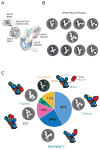
Figure 1. EM analysis of the conformational dynamics
(A) 3D reconstruction of the PI3KC3-C1 complex with the docked structures shown in a ribbon representation. (B) Representative class averages of the PI3KC3-C1 complex with the VPS34 HELCAT in range of different conformation states. (C) Percentage of well-resolved particles sorted into 2D class averages with a classical, V-shaped, WD, paddle and dislodged VPS34 HELCAT. Schematic and representative class averages of the PI3KC3-C1 complex showing the conformations of the VPS34 HELCAT.
Figure 2
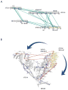
Figure 2. CX-MS analysis of the conformational dynamics
Pi3KC3-C1 was cross-linked with a lysine-specific bifunctional cross-linker, then fragmented by proteolysis, and cross-linked peptides were identified by mass spectrometry. (A) Inter-subunit cross-links between subunits of the PI3KC3-C1 complex. (B) Overview of cross-links observed within the PI3KC3-C1 complex corresponding to the model generated based on crystal structure of yeast complex I (PDB 5DFZ). The crosslinks are presented in red (> 38 Å) and blue (<38 Å), respectively. See also Table S1 and Movie S1.
Figure 3
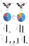
Figure 3. Leashing of the VPS34 HELCAT to VPS15
(A) Schematic of the PI3KC3-C1 complex showing leashed and unleashed construct of the VPS34 HELCAT. (B) Percentage of well-resolved particles sorted into 2D class averages with a classical, V-shaped, WD, paddle and dislodged VPS34 HELCAT in context of leashed and unleashed construct. (C) Quantification of PI(3)P generated by leashed and unleashed PI3KC3-C1 from PI and [γ-32 P] ATP. Samples were normalized to the activity of wt PI3KC3-C1 complex (D) Pho8Δ60 assay to monitor autophagy in yeast was carried out in rich (grey) or nitrogen starvation (black) media. Samples were normalized to the activity of Vps34 in rapamycin-treated cells. (E) Quantification of PI(3)P generated by fully assembled PI3KC3-C1 and as compared to isolated full-length VPS34. Data are represented as mean of three independent experiments +/− SD. See also Figure S1, Figure S2, Figure S3 and Figure S4.
Figure 4
Figure 4. PI3K complex assembly and activation model
Schematic of the PI3KC3-C1 complex showing the role of VPS15 in the assembly pathway and in regulating the dynamics and activity of the VPS34 HELCAT.

References

    1. Aebersold R, Mann M. Mass-spectrometric exploration of proteome structure and function. Nature. 2016;537:347–355. - PubMed
    1. Backer JM. The regulation and function of Class III PI3Ks: novel roles for Vps34. Biochem J. 2008;410:1–17. - PubMed
    1. Backer JM. The intricate regulation and complex functions of the Class III phosphoinositide 3-kinase Vps34. Biochem J. 2016;473:2251–2271. - PubMed
    1. Baskaran S, Carlson LA, Stjepanovic G, Young LN, Kim DJ, Grob P, Stanley RE, Nogales E, Hurley JH. Architecture and Dynamics of the Autophagic Phosphatidylinostol 3-Kinase Complex. eLife. 2014 Dec;9:3. doi: 10.7554/eLife.05115. - DOI - PMC - PubMed
    1. Chait BT, Cadene M, Olinares PD, Rout MP, Shi Y. Revealing Higher Order Protein Structure Using Mass Spectrometry. J Am Soc Mass Spectrom. 2016;27:952–965. - PMC - PubMed

MeSH terms

Substances