. 2017 Aug 3;67(3):361-373.e4.
doi: 10.1016/j.molcel.2017.06.034. Epub 2017 Jul 27.

AID Recognizes Structured DNA for Class Switch Recombination

Affiliations

AID Recognizes Structured DNA for Class Switch Recombination

Qi Qiao et al. Mol Cell. .

Abstract

Activation-induced cytidine deaminase (AID) initiates both class switch recombination (CSR) and somatic hypermutation (SHM) in antibody diversification. Mechanisms of AID targeting and catalysis remain elusive despite its critical immunological roles and off-target effects in tumorigenesis. Here, we produced active human AID and revealed its preferred recognition and deamination of structured substrates. G-quadruplex (G4)-containing substrates mimicking the mammalian immunoglobulin switch regions are particularly good AID substrates in vitro. By solving crystal structures of maltose binding protein (MBP)-fused AID alone and in complex with deoxycytidine monophosphate, we surprisingly identify a bifurcated substrate-binding surface that explains structured substrate recognition by capturing two adjacent single-stranded overhangs simultaneously. Moreover, G4 substrates induce cooperative AID oligomerization. Structure-based mutations that disrupt bifurcated substrate recognition or oligomerization both compromise CSR in splenic B cells. Collectively, our data implicate intrinsic preference of AID for structured substrates and uncover the importance of G4 recognition and oligomerization of AID in CSR.

Keywords: AID; APOBEC; CSR; G-quadruplex; activation-induced cytidine deaminase; class switch recombination; crystal structure.


Figures

Figure 1
Figure 1. Active Monomeric AID from Protein Engineering
(A) A schematic diagram of construct design with indicated mutation sites. More details are shown in Figure S1A. (B) Gel filtration chromatography of WT AID, AID.mono+CTT, AID.mono, and AID.mono after MBP removal. The measured molecular masses by multi-angle light scattering (MALS) and the theoretical molecular masses are shown. (C) In vitro deamination assay showing that both monomeric AID.mono and AID.mono+CTT had much higher activity than aggregated AID on ssDNA. Experiments used 0.1 μM AID and 1 μM DNA. (D) Rifampicin resistance (RifR) assay (Petersen-Mahrt et al., 2002) in E. coli KL16 and its uracil-DNA glycosylase (UDG)-deficient derivative BW310 (ung−/−), showing mutation frequencies by AID.WT and AID with mutations in AID.mono and AID.crystal. UDG removes the uracil base as a first step in base-excision repair following cytidine deamination, and its deficiency enhances AID-mediated mutation frequency. Data are represented as mean ± SD from 12 independent measurements. (E) CSR rescue of AID-deficient splenic B cells ex vivo by AID.WT and by AID with mutations in AID.mono and AID.crystal. Percent (%) CSR to IgG1 is the ratio between GFP+/IgG1+ cells (upper right quadrant) and total GFP+ cells (the two right quadrants). Data are represented as mean ± SD from three to six independent measurements. See also Figure S1.
Figure 2
Figure 2. Linear and G4 Structured Substrates
(A) Two types of AID substrates: one with multiple G-repeats and hotspots as in a mouse Sμ fragment and the other with a single G repeat and hotspot. The potential ability of these substrates to form either intramolecular or intermolecular G4 structure is illustrated. (B) G4 structure formation confirmed by the G4-specific dye N-methyl mesoporphyrin IX (NMM). LiCl is known to inhibit G4 structures. In total, 10 μM DNA and 20 μM NMM were used in each experiment. Heat denaturation was done by 95°C incubation for 10 min followed by flash cooling to eliminate DNA structures. Data are represented as mean ± SD from three independent measurements. (C) Separation of intermolecular G4 and linear fractions of single G-repeat substrate using a Superdex 75 gel filtration column. (D) Native gel showing the clear separation of linear and G4 structured substrate from Superdex 75 gel filtration purification in (C). See also Figure S2.
Figure 3
Figure 3. AID Preferentially Binds and Deaminates G4 Structured Substrates
(A) Electrophoretic mobility shift assay (EMSA) curves showing the significantly higher AID binding affinity of G4 fractions than linear fractions with identical primary sequences. (B) KD calculated by EMSA showing that ssDNA overhangs in G4 substrates are required for AID binding. Affinities increased with overhang length and plateaued at 5 nt. (C) EMSA showing that AID binds to RNA G4 similarly as to DNA G4. (D) In vitro deamination assays showing that AID has a higher deamination activity on G4 substrates than on linear substrates, with or without hotspots. Experiments used 0.1 μM AID and 1 μM DNA. (E) Competition assays showing that excess linear substrates did not compete with G4 substrates. Experiments used 1 μM AID, 1 μM substrate DNA, and up to 100 μM competitor ssDNA. Reaction time was 10 min. (F) In vitro deamination assay showing that Apobec3A and 3G did not exhibit G4 preference. Experiments used 0.1 μM APOBEC protein and 1 μM substrate DNA. (G) In vitro deamination assays showing that the peak activity of AID appears when the substrate nucleotide is at the third position 3′ to the G4 core. Experiments used 0.1 μM AID and 1 μM DNA. Reaction time was 10 min. (H) In vitro deamination assays showing that AID oligomerization causes clustered mutations. Experiments used 0.2 μM DNA and 0.2, 0.4, or 0.8 μM AID.mono. Reaction time was 2 min. Data in (A)–(C), (G), and (H) are represented as mean ± SD from three independent measurements. See also Figure S3.
Figure 4
Figure 4. Structures of AID and Its Complex with dCMP
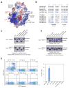
(A) Gel filtration chromatography with in-line MALS showing that AID.crystal binds G4 DNA in 2:1 ratio and branched DNA in 1:1 ratio. Measured and calculated molecular masses are labeled. (B) EMSA curves showing enhanced AID binding affinity for branched substrate with two overhangs (red), in comparison to that with one overhang (linear substrate, black) or no overhang (dsDNA, blue). Data are represented as mean ± SD from three independent measurements. (C) Ribbon diagram of human AID in rainbow color showing the secondary structures and catalytic residues near active site Zn2+. W, water. (D) Locations of F42, F141, and F145 mutated in AID.crystal on the face of the crystal structure opposite to the active site. (E) Surface charge distribution of the AID/dCMP structure and pocket prediction revealed a substrate binding channel that passes through the active site (green mesh). (F) Comparison between the AID-APOBEC3A hybrid AIDv (PDB: 5JJ4) and AID.crystal showed distinct surface charge distribution at the substrate channel. (G) Substrate dCMP in AID (E58A) catalytic center showing the interactions with surrounding residues. The E58 side chain is taken from the WT Apo-AID structure. (H) Alignment between Apo- and dCMP-bound AID structures showing the movement of R25 and N51 upon substrate recognition. (I) Mutations associated with the hyper-IgM syndrome mapped to AID structure. See also Figure S4 and Tables S1–S3.
Figure 5
Figure 5. AID Structures Revealed a Bifurcated Recognition Site for Two ssDNA Overhangs
(A) Surface charge distribution of AID structure revealed an additional positive patch away from the substrate-binding channel for recognition of an additional ssDNA. (B) Sequence alignment among different species of AID as well as human APOBECs, showing that the unique basic residue distribution in the substrate channel and the assistant patch is not conserved in APOBECs. (C) Mutations on positively charged residues in the substrate chain channel abolished deamination on substrates with either one or two ssDNA overhangs. The K34S/R77S/R107S mutant is a negative control on positively charged residues elsewhere. (D) Mutations on positively charged residues on the assistant patch impaired deamination on the substrate with two overhangs, without significantly affecting AID activity on that with one overhang. Experiments in (C) and (D) used 0.1 μM AID and 1 μM DNA. (E) Mutations on the assistant patch abolished CSR. The AIDv was also completely deficient in rescuing CSR. Notably, all AID constructs in this assay contain intact CTT, including AIDv. Data are represented as mean ± SD from three independent measurements. See also Figure S5.
Figure 6
Figure 6. AID Oligomerization and Structural Comparison with APOBECs
(A) Bio-layer interferometry by Blitz showing the kinetics of a hotspot containing G4 substrate binding by AID.mono (left) and AID.crystal (right). Much faster dissociation was observed for AID.crystal in comparison with AID.mono. (B) Bio-layer interferometry by Blitz showing the kinetics of non-hotspot G4 substrate binding by AID.mono (left) and AID.crystal (right). The much faster dissociation remained for AID.crystal. (C) The U-shaped substrate-binding channel observed in A3A-ssDNA complex structure (PDB: 5SWW). (D) AID surface with the U-shaped substrate from in A3A, showing steric clash. (E) A structure model of AID in complex with the AGCTT ssDNA. (F) A schematic diagram suggesting that a U-shaped substrate channel in A3A and A3B may not support structured substrate recognition. See also Figure S6.
Figure 7
Figure 7
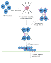
A Model of G4-Structure-Mediated AID Recruitment and Oligomerization that Create Mutation Clusters and DSB

Similar articles

Cited by

References

    1. Adams PD, Afonine PV, Bunkóczi G, Chen VB, Davis IW, Echols N, Headd JJ, Hung LW, Kapral GJ, Grosse-Kunstleve RW, et al. PHENIX: a comprehensive Python-based system for macromolecular structure solution. Acta Crystallogr D Biol Crystallogr. 2010;66:213–221. - PMC - PubMed
    1. Bardin C, Leroy JL. The formation pathway of tetramolecular G-quadruplexes. Nucleic Acids Res. 2008;36:477–488. - PMC - PubMed
    1. Basu U, Chaudhuri J, Alpert C, Dutt S, Ranganath S, Li G, Schrum JP, Manis JP, Alt FW. The AID antibody diversification enzyme is regulated by protein kinase A phosphorylation. Nature. 2005;438:508–511. - PubMed
    1. Bransteitter R, Pham P, Scharff MD, Goodman MF. Activation-induced cytidine deaminase deaminates deoxycytidine on single-stranded DNA but requires the action of RNase. Proc Natl Acad Sci USA. 2003;100:4102–4107. - PMC - PubMed
    1. Chambers VS, Marsico G, Boutell JM, Di Antonio M, Smith GP, Balasubramanian S. High-throughput sequencing of DNA G-quadruplex structures in the human genome. Nat Biotechnol. 2015;33:877–881. - PubMed

MeSH terms