

Endotoxemia induces lung-brain coupling and multi-organ injury following cerebral ischemia-reperfusion
- PMID: 28757259
- PMCID: PMC5612886
- DOI: 10.1016/j.expneurol.2017.07.016
Endotoxemia induces lung-brain coupling and multi-organ injury following cerebral ischemia-reperfusion
Abstract
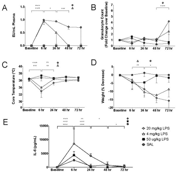
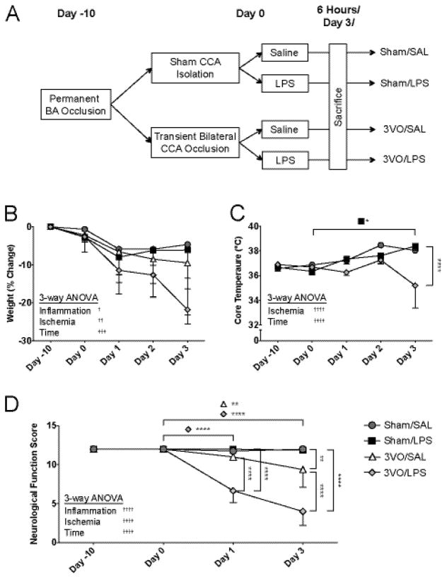
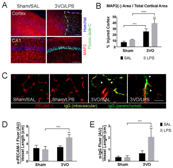
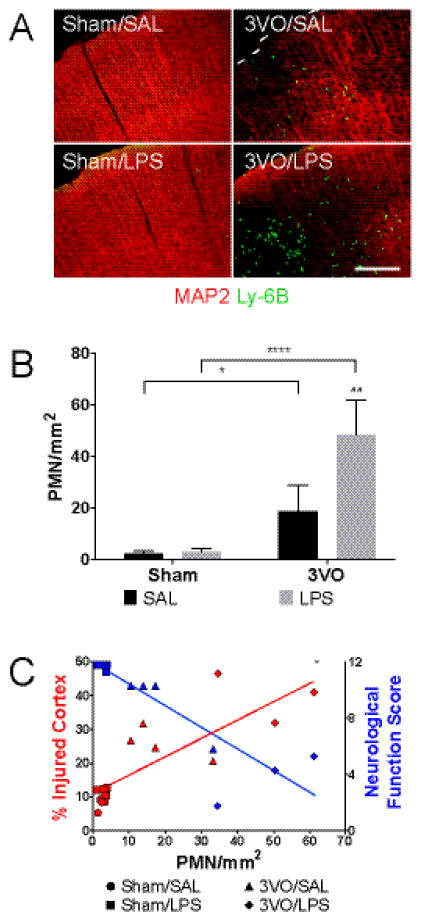
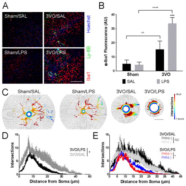
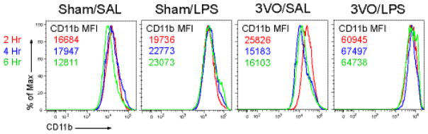
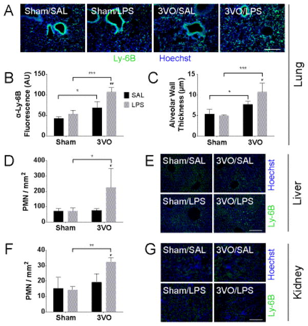
Post-ischemic neurodegeneration remains the principal cause of mortality following cardiac resuscitation. Recent studies have implicated gastrointestinal ischemia in the sepsis-like response associated with the post-cardiac arrest syndrome (PCAS). However, the extent to which the resulting low-grade endotoxemia present in up to 86% of resuscitated patients affects cerebral ischemia-reperfusion injury has not been investigated. Here we report that a single injection of low-dose lipopolysaccharide (50μg/kg, IP) delivered after global cerebral ischemia (GCI) induces blood-brain barrier permeability, microglial activation, cortical injury, and functional decline in vivo, compared to ischemia alone. And while GCI was sufficient to induce neutrophil (PMN) activation and recruitment to the post-ischemic CNS, minimal endotoxemia exhibited synergistic effects on markers of systemic inflammation including PMN priming, lung damage, and PMN burden within the lung and other non-ischemic organs including the kidney and liver. Our findings predict that acute interventions geared towards blocking the effects of serologically occult endotoxemia in survivors of cardiac arrest will limit delayed neurodegeneration, multi-organ dysfunction and potentially other features of PCAS. This work also introduces lung-brain coupling as a novel therapeutic target with broad effects on innate immune priming and post-ischemic neurodegeneration following cardiac arrest and related cerebrovascular conditions.
Keywords: Blood-brain barrier; Endotoxin; Global cerebral ischemia; Ischemia-reperfusion injury; Microglia; Neurodegeneration; Neuroinflammation; Neutrophil; Post-cardiac arrest syndrome; Systemic inflammation.
Copyright © 2017 Elsevier Inc. All rights reserved.
Conflict of interest statement
None.
Figures
Similar articles
-
The post-cardiac arrest syndrome: A case for lung-brain coupling and opportunities for neuroprotection.J Cereb Blood Flow Metab. 2019 Jun;39(6):939-958. doi: 10.1177/0271678X19835552. Epub 2019 Mar 13. J Cereb Blood Flow Metab. 2019. PMID: 30866740 Free PMC article. Review.
-
Lung-Derived SOD3 Attenuates Neurovascular Injury After Transient Global Cerebral Ischemia.J Am Heart Assoc. 2019 May 7;8(9):e011801. doi: 10.1161/JAHA.118.011801. J Am Heart Assoc. 2019. PMID: 31030600 Free PMC article.
-
Role of nitric oxide in hepatic ischemia-reperfusion with endotoxemia.J Inflamm. 1995-1996;46(3):144-54. J Inflamm. 1995. PMID: 8844495
-
Lung SOD3 limits neurovascular reperfusion injury and systemic immune activation following transient global cerebral ischemia.J Stroke Cerebrovasc Dis. 2020 Sep;29(9):104942. doi: 10.1016/j.jstrokecerebrovasdis.2020.104942. Epub 2020 May 14. J Stroke Cerebrovasc Dis. 2020. PMID: 32807413 Free PMC article.
-
Role of microglia under cardiac and cerebral ischemia/reperfusion (I/R) injury.Metab Brain Dis. 2018 Aug;33(4):1019-1030. doi: 10.1007/s11011-018-0232-4. Epub 2018 Apr 14. Metab Brain Dis. 2018. PMID: 29656335 Review.
Cited by
-
Characterization of neutrophil-neuronal co-cultures to investigate mechanisms of post-ischemic immune-mediated neurotoxicity.J Neurosci Methods. 2020 Jul 15;341:108782. doi: 10.1016/j.jneumeth.2020.108782. Epub 2020 May 20. J Neurosci Methods. 2020. PMID: 32445795 Free PMC article.
-
Chronic stress and corticosterone exacerbate alcohol-induced tissue injury in the gut-liver-brain axis.Sci Rep. 2021 Jan 12;11(1):826. doi: 10.1038/s41598-020-80637-y. Sci Rep. 2021. PMID: 33436875 Free PMC article.
-
Adaptive Immune Cells Link Microbial Metabolites to Stroke Recovery.J Neurosci. 2020 Jul 8;40(28):5344-5346. doi: 10.1523/JNEUROSCI.0634-20.2020. J Neurosci. 2020. PMID: 32641452 Free PMC article. No abstract available.
-
The post-cardiac arrest syndrome: A case for lung-brain coupling and opportunities for neuroprotection.J Cereb Blood Flow Metab. 2019 Jun;39(6):939-958. doi: 10.1177/0271678X19835552. Epub 2019 Mar 13. J Cereb Blood Flow Metab. 2019. PMID: 30866740 Free PMC article. Review.
-
Propofol inhibits neuroinflammation and metabolic reprogramming in microglia in vitro and in vivo.Front Pharmacol. 2023 Jun 13;14:1161810. doi: 10.3389/fphar.2023.1161810. eCollection 2023. Front Pharmacol. 2023. PMID: 37383725 Free PMC article.
References
-
- Abella BS, Zhao D, Alvarado J, Hamann K, Vanden Hoek TL, Becker LB. Intra-arrest cooling improves outcomes in a murine cardiac arrest model. Circulation. 2004;109:2786–2791. - PubMed
-
- Abraham E. Neutrophils and acute lung injury. Critical care medicine. 2003;31:S195–199. - PubMed
-
- Adrie C, Adib-Conquy M, Laurent I, Monchi M, Vinsonneau C, Fitting C, et al. Successful cardiopulmonary resuscitation after cardiac arrest as a “sepsis-like” syndrome. Circulation. 2002;106:562–568. - PubMed
-
- Adrie C, Laurent I, Monchi M, Cariou A, Dhainaou JF, Spaulding C. Postresuscitation disease after cardiac arrest: A sepsis-like syndrome? Current opinion in critical care. 2004;10:208–212. - PubMed
-
- Annane D, Sharshar T. Cognitive decline after sepsis. Lancet Respir Med. 2015;3:61–69. - PubMed
Publication types
MeSH terms
Substances
Grants and funding
LinkOut - more resources
Full Text Sources
Other Literature Sources
