

Pathophysiology of Eosinophilic Esophagitis
- PMID: 28757265
- PMCID: PMC5787048
- DOI: 10.1053/j.gastro.2017.06.065
Pathophysiology of Eosinophilic Esophagitis
Abstract
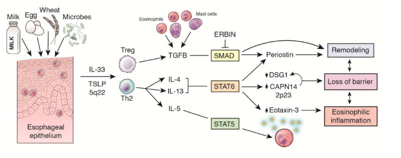
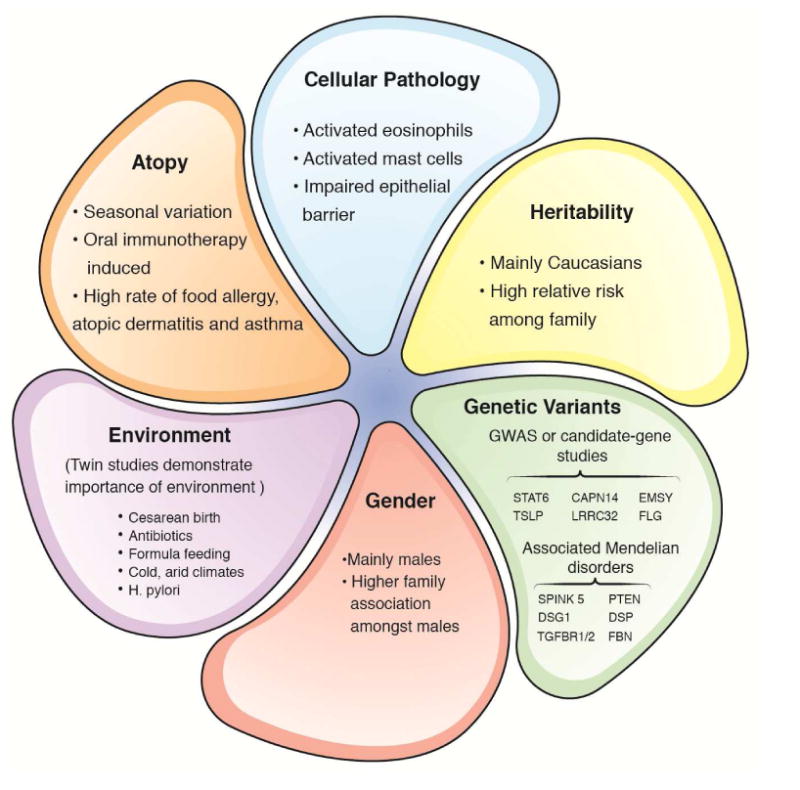
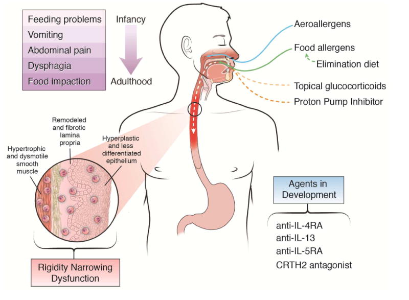
Eosinophilic esophagitis is an emerging disease that is distinguished from gastroesophageal reflux disease by the expression of a unique esophageal transcriptome and the interplay of early life environmental factors with distinct genetic susceptibility elements at 5q22 (TSLP) and 2p23 (CAPN14). Rare genetic syndromes have uncovered the contribution of barrier disruption, mediated in part by defective desmosomes and dysregulated transforming growth factor beta production and signaling, to eosinophilic esophagitis pathophysiology. Experimental modeling has defined a cooperative role of activated eosinophils, mast cells, and the cytokines IL-5 and IL-13, mediated by allergic sensitization to multiple foods. Understanding these processes is opening the way to better treatment based on disrupting allergic inflammatory and type 2 cytokine-mediated responses, including anti-cytokine therapeutics and dietary therapy.
Keywords: Allergy; Desmosome; Genetics; Inflammation.
Copyright © 2018 AGA Institute. Published by Elsevier Inc. All rights reserved.
Figures
References
-
- Dellon ES, Gonsalves N, Hirano I, et al. ACG clinical guideline: Evidenced based approach to the diagnosis and management of esophageal eosinophilia and eosinophilic esophagitis (EoE) Am J Gastroenterol. 2013;108:679–92. - PubMed
-
- Liacouras CA, Furuta GT, Hirano I, et al. Eosinophilic esophagitis: updated consensus recommendations for children and adults. J Allergy Clin Immunol. 2011;128:3–20. - PubMed
-
- Collins MH. Histopathologic features of eosinophilic esophagitis. Gastroenterology Clinics of North America. 2014;43:257–68. - PubMed
Publication types
MeSH terms
Substances
Supplementary concepts
Grants and funding
LinkOut - more resources
Full Text Sources
Other Literature Sources
Medical
