Review
. 2017 Nov;17(11):679-690.
doi: 10.1038/nri.2017.74. Epub 2017 Jul 31.

Mechanical forces in the immune system

Affiliations
Review

Mechanical forces in the immune system

Morgan Huse. Nat Rev Immunol. 2017 Nov.

Abstract

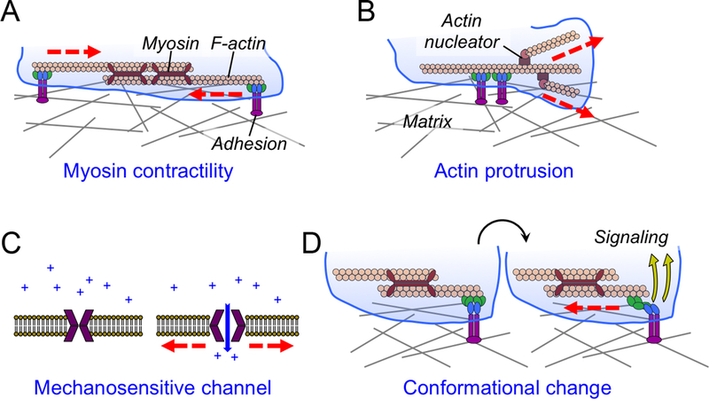
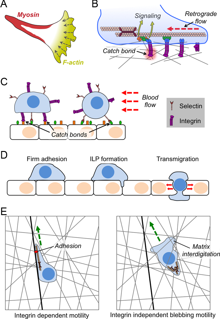
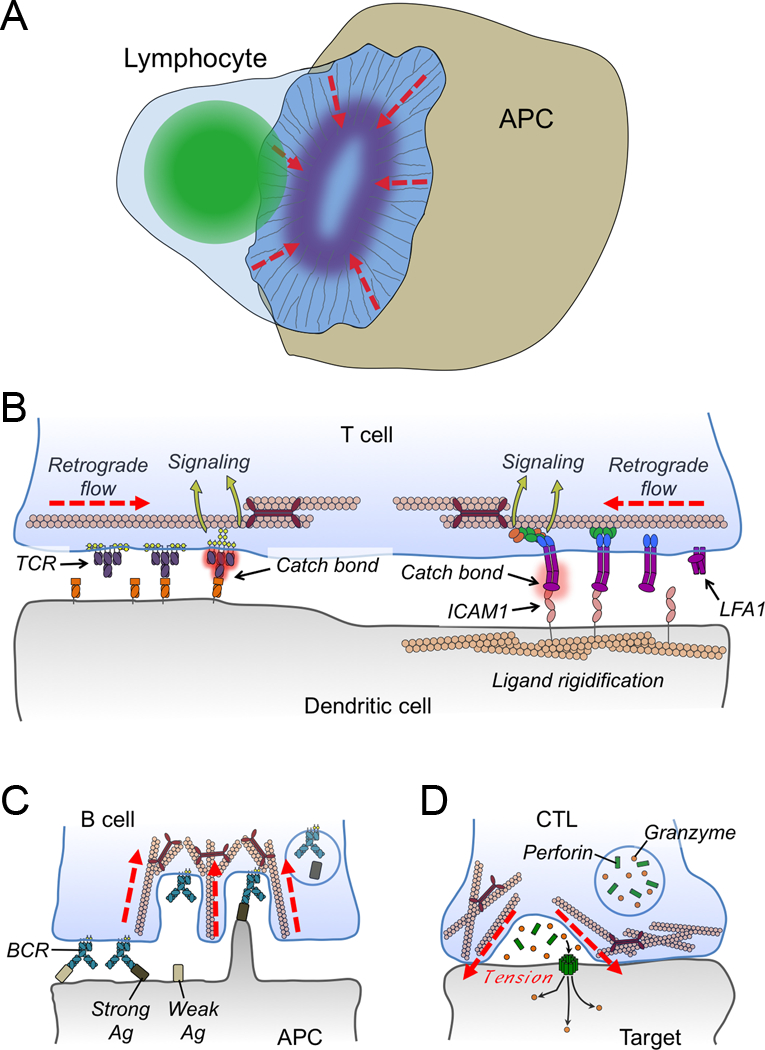
Leukocytes can completely reorganize their cytoskeletal architecture within minutes. This structural plasticity, which facilitates their migration and communicative function, also enables them to exert a substantial amount of mechanical force against the extracellular matrix and the surfaces of interacting cells. In recent years, it has become increasingly clear that these forces have crucial roles in immune cell activation and subsequent effector responses. Here, I review our current understanding of how mechanical force regulates cell-surface receptor activation, cell migration, intracellular signalling and intercellular communication, highlighting the biological ramifications of these effects in various immune cell types.


Figures

Figure 1.
Figure 1.. Force exertion and mechanotransduction.
(A-B) Cells exert forces against their environment through myosin contractility (A) and F-actin-based protrusion (B). (C) The mechanical gating of a plasma membrane channel by membrane tension. (D) Schematic diagram showing mechanotransduction induced by force dependent conformational change (blue and green components of the adhesion). In all figures, myosin and F-actin are shown as maroon, rabbit-eared oligomers and peach-colored polymers, respectively. Integrin-mediated adhesions are shown in purple, blue, and green. Forces are indicated by red lines.
Figure 2.
Figure 2.. Mechanical forces in cell migration.
(A) Image depicting the “hand mirror” morphology adopted by leukocytes migrating in two dimensions. (B) Diagram schematizing integrin catch bond and molecular clutch formation during cell migration. Integrins are shown in purple with associated focal adhesion proteins in blue, green, and orange. (C) Selectin and integrin catch bonds mediate leukocyte rolling (right) and firm adhesion (left) to endothelial walls under shear stress. (D) Transendothelial migration is initiated by the formation of an invadosome like protrusion (ILP), followed by the extrusion of the entire cell through the resulting pore. Force exertion is primarily seen in the transmigration step. (E) Schematic diagram contrasting adhesion dependent lamellipodial migration (left) and adhesion independent blebbing migration (right). Matrix is indicated by gray lines, and a fiber containing integrin ligands as a black line. Green arrows indicate the direction of motion. In B-D, red lines indicate force.
Figure 3.
Figure 3.. Mechanical forces in immune cell-cell interactions.
(A) Diagram schematizing the lymphocyte immunological synapse. Actin fibers are shown as thin gray lines, the lymphocyte nucleus is green, and integrin clustering is indicated by the purple oval. Dashed red lines indicate the direction of retrograde F-actin flow. (B) Retrograde flow is responsible for driving centripetal motion of TCRs (left) and LFA1 (right), which is thought to contribute to the formation of catch bonds, receptor conformational change, and subsequent mechanotransduction. DCs restrain the movement of cell surface ICAM1 to enhance LFA1 binding and signaling. (C) Myosin-based contractility enables B cells to selectively internalize high affinity antigen (Ag). (D) Synaptic forces enhance CTL-mediated killing by applying tension to the target cell, thereby potentiating perforin pore formation in the target cell membrane. In B-D, red lines denote forces.

References

    1. Iskratsch T, Wolfenson H & Sheetz MP Appreciating force and shape-the rise of mechanotransduction in cell biology. Nat Rev Mol Cell Biol 15, 825–833, doi:10.1038/nrm3903 (2014). - DOI - PMC - PubMed
    1. Vicente-Manzanares M, Ma X, Adelstein RS & Horwitz AR Non-muscle myosin II takes centre stage in cell adhesion and migration. Nat Rev Mol Cell Biol 10, 778–790, doi:nrm2786 [pii] 10.1038/nrm2786 (2009). - DOI - PMC - PubMed
    1. Smith A et al. The role of the integrin LFA-1 in T-lymphocyte migration. Immunol Rev 218, 135–146, doi:10.1111/j.1600-065X.2007.00537.x (2007). - DOI - PubMed
    1. Matsumura F Regulation of myosin II during cytokinesis in higher eukaryotes. Trends Cell Biol 15, 371–377, doi:10.1016/j.tcb.2005.05.004 (2005). - DOI - PubMed
    1. Hui KL, Balagopalan L, Samelson LE & Upadhyaya A Cytoskeletal forces during signaling activation in Jurkat T-cells. Mol Biol Cell 26, 685–695, doi:10.1091/mbc.E14-03-0830 (2015). - DOI - PMC - PubMed