Review
. 2017:2017:9193831.
doi: 10.1155/2017/9193831. Epub 2017 Jul 5.

Peripheral Exophytic Oral Lesions: A Clinical Decision Tree

Affiliations
Review

Peripheral Exophytic Oral Lesions: A Clinical Decision Tree

Hamed Mortazavi et al. Int J Dent. 2017.

Abstract

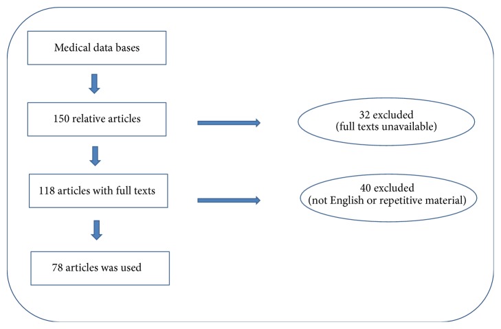
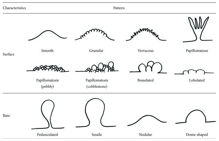
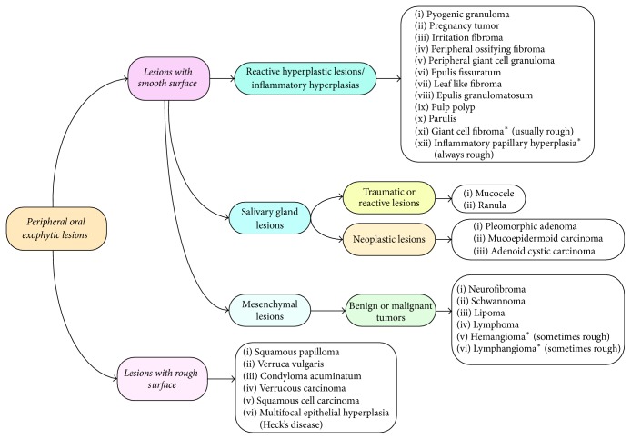
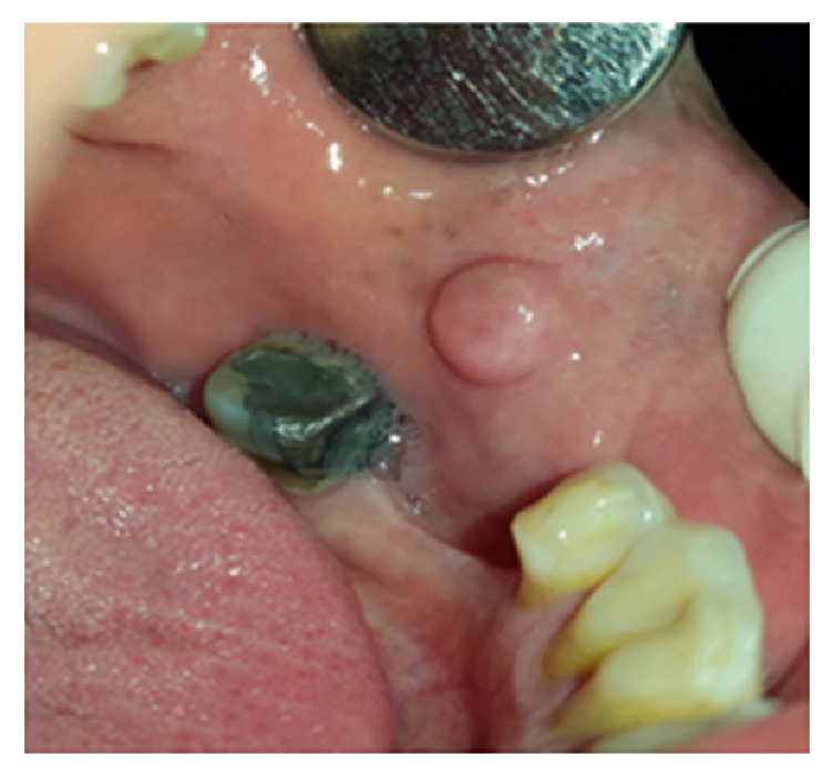
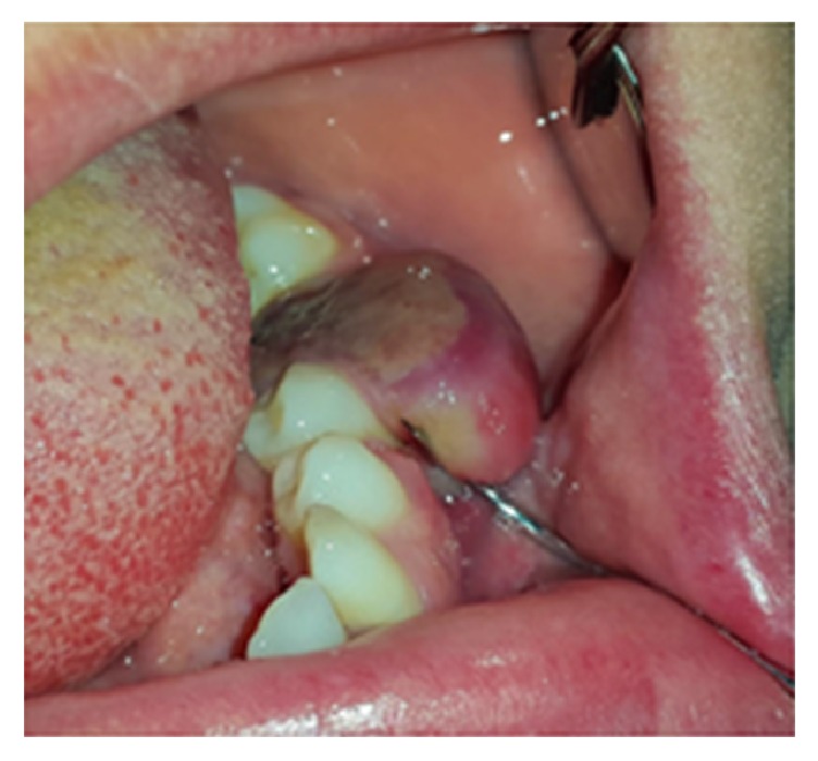
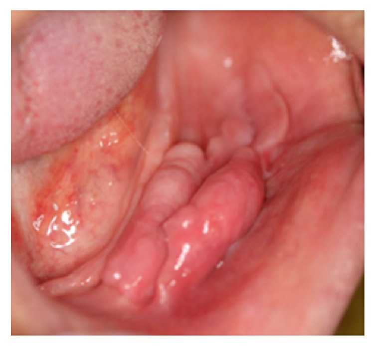
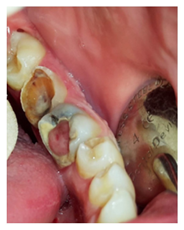
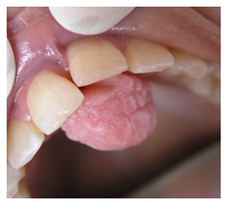
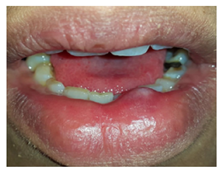
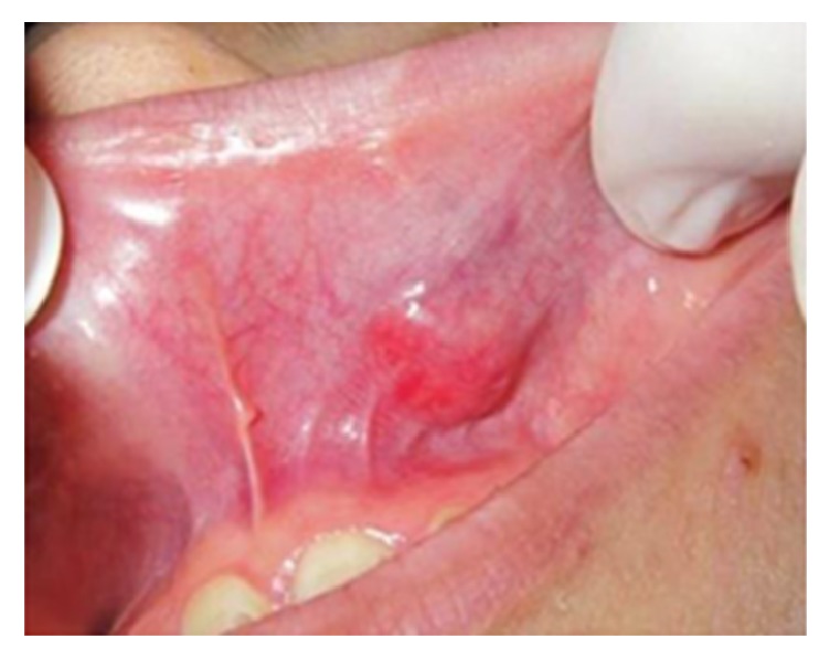
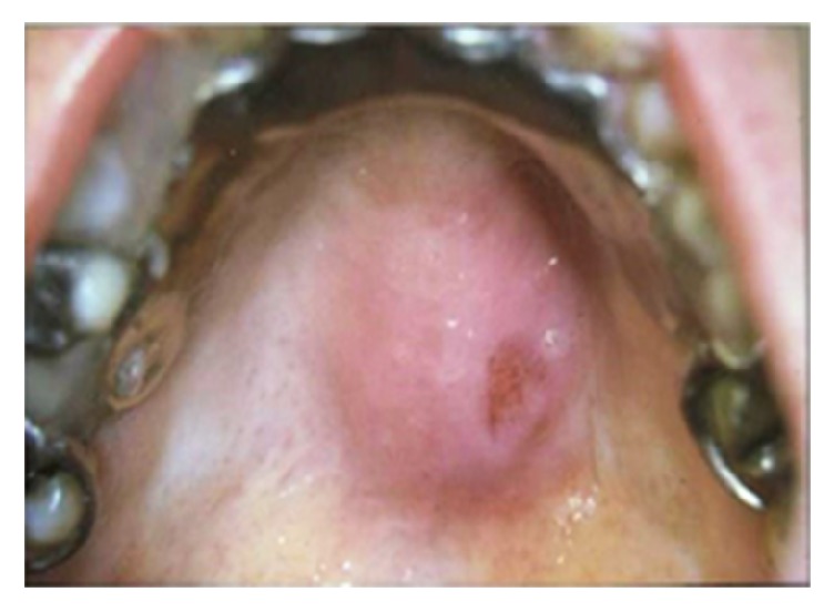
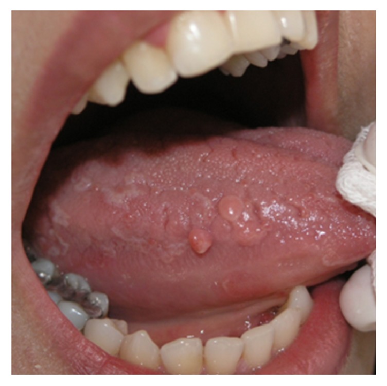
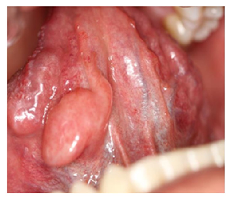
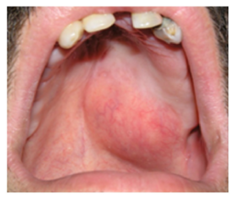
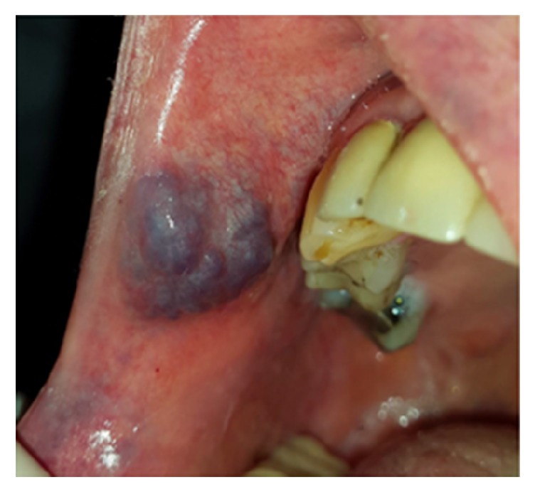
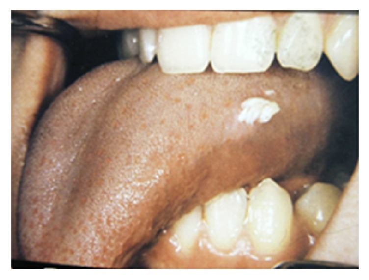
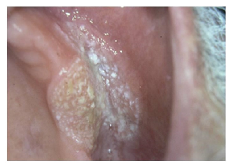
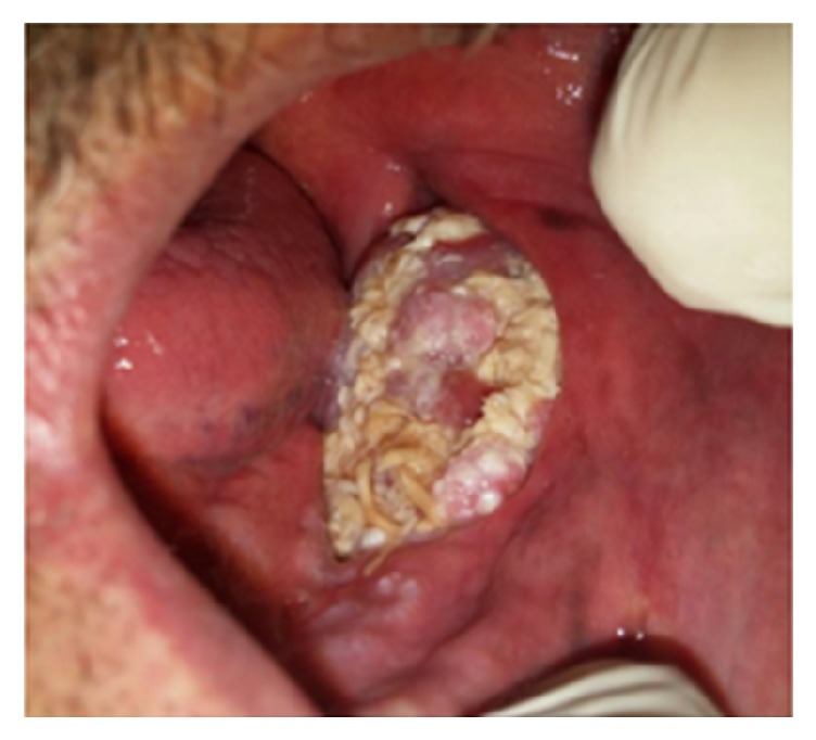
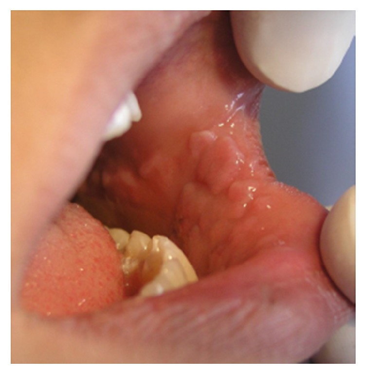
Diagnosis of peripheral oral exophytic lesions might be quite challenging. This review article aimed to introduce a decision tree for oral exophytic lesions according to their clinical features. General search engines and specialized databases including PubMed, PubMed Central, Medline Plus, EBSCO, Science Direct, Scopus, Embase, and authenticated textbooks were used to find relevant topics by means of keywords such as "oral soft tissue lesion," "oral tumor like lesion," "oral mucosal enlargement," and "oral exophytic lesion." Related English-language articles published since 1988 to 2016 in both medical and dental journals were appraised. Upon compilation of data, peripheral oral exophytic lesions were categorized into two major groups according to their surface texture: smooth (mesenchymal or nonsquamous epithelium-originated) and rough (squamous epithelium-originated). Lesions with smooth surface were also categorized into three subgroups according to their general frequency: reactive hyperplastic lesions/inflammatory hyperplasia, salivary gland lesions (nonneoplastic and neoplastic), and mesenchymal lesions (benign and malignant neoplasms). In addition, lesions with rough surface were summarized in six more common lesions. In total, 29 entities were organized in the form of a decision tree in order to help clinicians establish a logical diagnosis by a stepwise progression method.


Figures

Figure 1
Figure 1
Flowchart for choosing eligible articles.
Figure 2
Figure 2
Schematic view of surface and base characteristics of oral exophytic lesions.
Figure 3
Figure 3
Decision tree for peripheral oral exophytic lesions.
Figure 4
Figure 4
Pyogenic granuloma as a sessile lesion on mandibular labial gingivae with an ulcerated smooth surface.
Figure 5
Figure 5
Irritation fibroma on the buccal mucosa with a smooth surface and dome-shaped base.
Figure 6
Figure 6
PGCG with an ulcerated, smooth surface and purplish color located buccolingually on the left mandibular ridge.
Figure 7
Figure 7
Massive epulis fissuratum presenting as an exophytic lesion with smooth surface associated with an ill-fit mandibular denture.
Figure 8
Figure 8
Pulp polyp associated with carious first mandibular molar with smooth surface and sessile base.
Figure 9
Figure 9
Pedunculated lesion of giant cell fibroma with granular surface on the palatal gingivae of maxillary central incisors.
Figure 10
Figure 10
Nodular mucocele of the lower lip with smooth surface.
Figure 11
Figure 11
Pleomorphic adenoma involving upper lip presented as a dome-shaped exophytic lesion with smooth surface.
Figure 12
Figure 12
Mucoepidermoid carcinoma of the palate, presented as an exophytic lesion with smooth, ulcerated surface, and nodular base.
Figure 13
Figure 13
Sessile-based and smooth-surfaced exophytic lesions of neurofibroma involving dorsal and lateral border of the tongue.
Figure 14
Figure 14
A dome-shaped schwannoma on the ventral surface of the tongue with smooth surface.
Figure 15
Figure 15
Lymphoma presented as a nodular exophytic lesion with smooth surface on the palate.
Figure 16
Figure 16
Lobulated hemangioma involving the right upper lip.
Figure 17
Figure 17
Squamous papilloma involving lateral border of the tongue with a sessile base and papillomatous surface.
Figure 18
Figure 18
Broad-based verrucous carcinoma involving maxillary vestibular mucosa, with verrucous surface.
Figure 19
Figure 19
Exophytic SCC with a verrucous, necrotic, and ulcerative surface, extended from vestibule of the mandible to floor of the mouth.
Figure 20
Figure 20
Multifocal epithelial hyperplasia presented as numerous exophytic lesions with flat surface distributed in the buccal mucosa and lower lip.

References

    1. Wood N. K., Goaz P. W. Differential Diagnosis of Oral And Maxillofacial Lesions. 5th Edition. St. Louis, Mo, USA: Mosby; 1997.
    1. Santosh A. B. R., Boyd D., Laxminarayana K. K. Proposed clinico-pathological classification for oral exophytic lesions. Journal of Clinical and Diagnostic Research. 2015;9(9):ZE01–ZE08. doi: 10.7860/JCDR/2015/12662.6468. - DOI - PMC - PubMed
    1. Zain R. B., Ikeda N., Razak I. A., et al. A national epidemiological survey of oral mucosal lesions in Malaysia. Community Dentistry and Oral Epidemiology. 1997;25(5):377–383. doi: 10.1111/j.1600-0528.1997.tb00959.x. - DOI - PubMed
    1. Glick M. Burket's Oral Medicine. 12th edition. People's medical publishing house; 2015. 147-172, 175-188, 236-237.
    1. Bermejo-Fenoll A., López-Jornet P. Differential diagnosis of exophytic lesions of soft oral tissue. Medicina Oral, Patologia Oral y Cirugia Bucal. 2005;10(5):470–471. - PubMed

LinkOut - more resources