. 2015 Dec 1;6(12):7097-7104.
doi: 10.1039/c5sc02467f. Epub 2015 Sep 11.

Design, preparation, and selection of DNA-encoded dynamic libraries

Affiliations

Design, preparation, and selection of DNA-encoded dynamic libraries

Gang Li et al. Chem Sci. .

Abstract

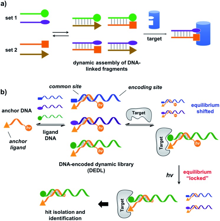
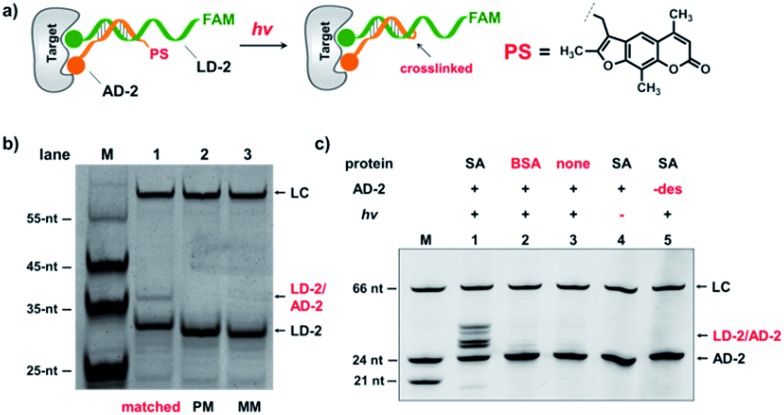
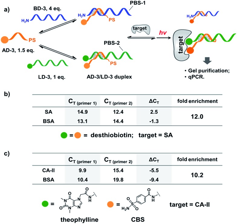
We report a method for the preparation and selection of DNA-encoded dynamic libraries (DEDLs). The library is composed of two sets of DNA-linked small molecules that are under dynamic exchange through DNA hybridization. Addition of the protein target shifted the equilibrium, favouring the assembly of high affinity bivalent binders. Notably, we introduced a novel locking mechanism to stop the dynamic exchange and "freeze" the equilibrium, thereby enabling downstream hit isolation and decoding by PCR amplification and DNA sequencing. Our DEDL approach has circumvented the limitation of library size and realized the analysis and selection of large dynamic libraries. In addition, this method also eliminates the requirement for modified and immobilized target proteins.


Figures

Fig. 1
Fig. 1. (a) Previous work: two sets of DNA-linked fragments form dynamically exchanging duplexes; addition of the target enriches high affinity duplexes.,, (b) DNA-encoded dynamic library (DEDL) (this work): an anchor DNA forms dynamic duplexes with multiple ligand DNA strands, forming the library. Adding the target shifts the equilibrium, favouring the formation of high affinity binders. A photo-crosslinker in the anchor DNA locks the shifted equilibrium under irradiation. Crosslinked binders can then be isolated for hit identification via PCR amplification and DNA sequencing.
Fig. 2
Fig. 2. (a) Two complementary DNA strands conjugated to biotin, desthiobiotin, or iminobiotin and FAM/DABCYL groups were mixed with the target SA (i), with BSA (ii), with no protein (iii), or with one ligand omitted (iv). (b) Structures of the small molecule ligands. (c) Fluorescence quenching results. FAM fluorescence values were measured and normalized to (iii). Left panel: with biotin; middle panel: with desthiobiotin; right panel: with iminobiotin. (d) Structure of the raloxifene ligand. (e) Fluorescence quenching results of the raloxifene–ER system. ER was used as the target in (i) and (iv). DNA: 200 nM each; protein: 400 nM. The DNA and protein were incubated at 30 °C for 1 h before measurement using a fluorophotometer. Excitation: 494 nm; emission: 522 nm. Error bars (standard deviation, SD) are based on three replicates of each experiment.
Fig. 3
Fig. 3. (a) Two desthiobiotin-labelled DNA strands conjugated to FAM or DABCYL were mixed with SA or BSA in different orders or at different temperatures; fluorescence decreases were then measured. (b) Left panel: data from different mixing orders. Right panel: data from different temperatures; SA was used as the target except in 5 where BSA was used as a negative control. The experimental conditions were the same as those for Fig. 2 except for the mixing order and temperature. Error bars (standard deviation, SD) are based on three replicates of each experiment.
Fig. 4
Fig. 4. Verification of the target-induced equilibrium shift, determined by a fluorescence decrease. Fluorescence values were normalized to the “no protein” experiment. AD-1 and LD-1: 200 nM; BD-1: 1.6 μM; proteins: 400 nM. The experimental procedures were the same as those for Fig. 2. Error bars (standard deviation, SD) are based on three replicates of each experiment.
Fig. 5
Fig. 5. (a) Psoralen (PS) was chosen to lock the equilibrium in the DEDL. (b) Three sets of AD-2/LD-2 were mixed, irradiated, and analysed by denaturing electrophoresis. PM: partially mismatched (2-base mismatch); MM: fully mismatched; LC: a 66-nt DNA loading control. (c) 5′-Desthiobiotin-labeled LD-2 and AD-2 were mixed, irradiated under different conditions, and then analysed by denaturing electrophoresis (18% TBE–urea denaturing PAGE). Lane 1: with SA; lane 2: with BSA; lane 3: no protein added; lane 4: no irradiation; lane 5: no desthiobiotin on AD-2. AD-2: 300 nM; LD-2: 200 nM; irradiation: 365 nm for 30 s at 30 °C using a UV LED point light system; short irradiation reduces non-specific crosslinking in the background. M: marker; –des: no desthiobiotin.
Fig. 6
Fig. 6. (a) PS-based photo-crosslinking locks the shifted equilibrium for the subsequent hit isolation and qPCR analysis. After gel-purification of the crosslinked duplexes, qPCR was performed to determine the C T values and to calculate the fold enrichment of the AD-3/LD-3 duplex. (b) Results of the bivalent desthiobiotin–SA system. (c) Results of the theophylline/CBS–CA-II system. ΔC T = C T (primer 1)C T (primer 2). AD-3: 300 nM; LD-3: 200 nM; primers: 200 nM. The experimental procedure was the same as that for Fig. 5. See the ESI for details.
Fig. 7
Fig. 7. (a) Components of the model libraries. Libraries were selected against SA with the same procedure as that for Fig. 6, except Sanger sequencing was used. (b) Sequencing results; left panels: after SA selection; right panels: control selection without SA. LD-4: 200 nM; BD-4: 800 nM (total); AD-4: 300 nM; SA: 400 nM. See the ESI for details.
Fig. 8
Fig. 8. (a) Components of the 1025-member library. The library was selected against SA with the same procedure as that for Fig. 6, except Illumina® sequencing was used. (b) Plot of the fold enrichment versus sequence count after the target selection. Fold enrichment = (post-target selection fraction)/(post-control selection fraction). (c) Plot of sequence count ratios after the control selection (no protein added) versus count ratios after the target selection (with SA). Sequence ratio = (sequence count)/(total sequence count of the library). Each dot represents the DNA sequence corresponding to a library member. The “hit” containing the desired LD-5 codon is highlighted in red. LD-5: 0.19 nM; BD-5: 200 nM (total); AD-5: 300 nM; SA: 400 nM. The fold enrichments for the low-count library members vary widely due to statistical under-sampling. See the ESI for more details on the experimental procedure, data analysis and further discussion of the sequencing results.

References

    1. Corbett P. T., Leclaire J., Vial L., West K. R., Wietor J.-L., Sanders J. K., Otto S. Chem. Rev. 2006;106:3652–3711. - PubMed
    1. Cougnon F. B., Sanders J. K. Acc. Chem. Res. 2011;45:2211–2221. - PubMed
    1. Jin Y., Yu C., Denman R. J., Zhang W. Chem. Soc. Rev. 2013;42:6634–6654. - PubMed
    1. Meyer C. D., Joiner C. S., Stoddart J. F. Chem. Soc. Rev. 2007;36:1705–1723. - PubMed
    1. Mondal M., Hirsch A. K. Chem. Soc. Rev. 2015;44:2455–2488. - PubMed