

Endophilin2 Interacts with GluA1 to Mediate AMPA Receptor Endocytosis Induced by Oligomeric Amyloid- β
- PMID: 28758034
- PMCID: PMC5516760
- DOI: 10.1155/2017/8197085
Endophilin2 Interacts with GluA1 to Mediate AMPA Receptor Endocytosis Induced by Oligomeric Amyloid- β
Abstract
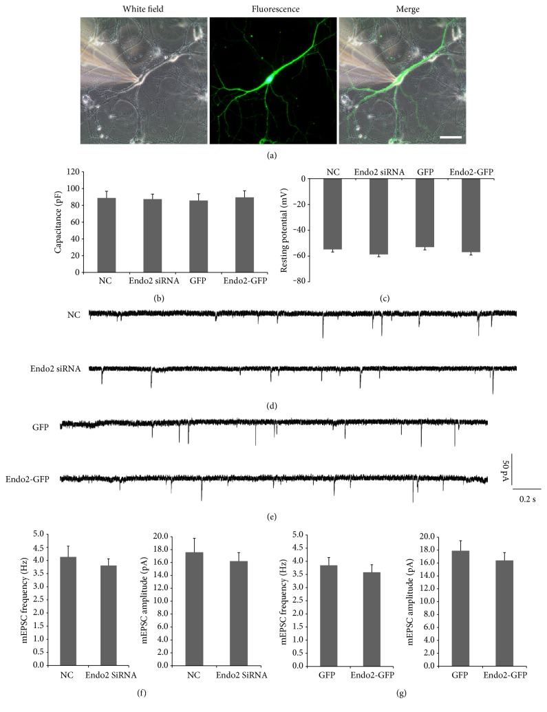
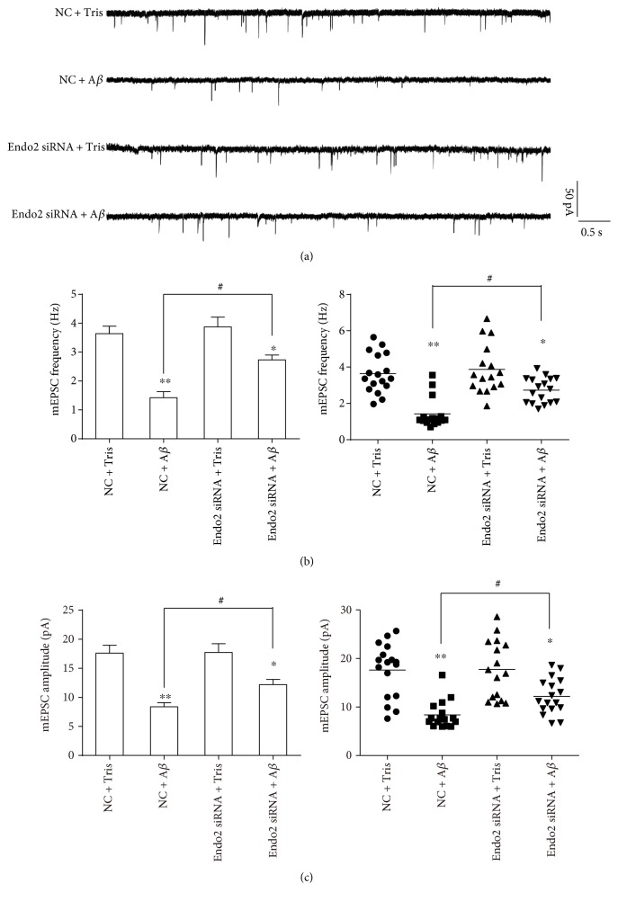
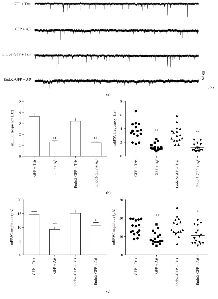
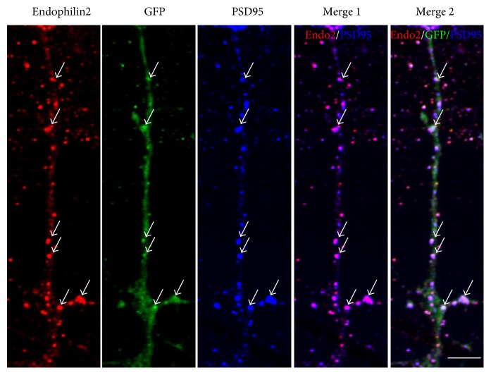
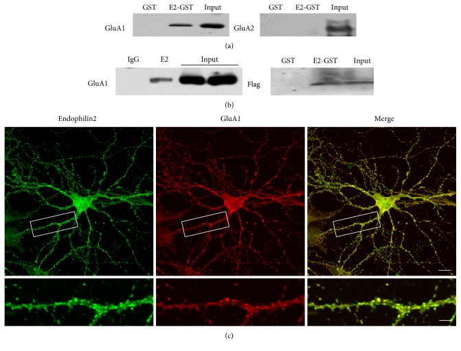
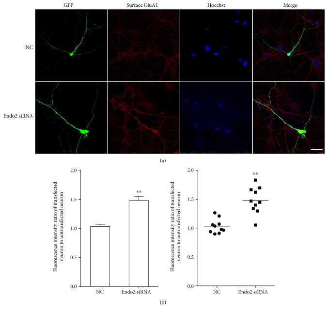
Amyloid-β (Aβ) plays an important role in Alzheimer's disease (AD), as oligomeric Aβ induces loss of postsynaptic AMPA receptors (AMPARs) leading to cognitive deficits. The loss of postsynaptic AMPARs is mediated through the clathrin-dependent endocytosis pathway, in which endophilin2 is one of the important regulatory proteins. Endophilin2, which is enriched in both the pre- and postsynaptic membrane, has previously been reported to be important for recycling of synaptic vesicles at the presynaptic membrane. However, the role of endophilin2 in oligomeric Aβ-induced postsynaptic AMPAR endocytosis is not well understood. In this study, we show that endophilin2 does not affect constitutive AMPAR endocytosis. Endophilin2 knockdown, but not overexpression, resisted oligomeric Aβ-induced AMPAR dysfunction. Moreover, endophilin2 colocalized and interacted with GluA1, a subunit of AMPAR, to regulate oligomeric Aβ-induced AMPAR endocytosis. Thus, we have determined a role of endophilin2 in oligomeric Aβ-induced postsynaptic AMPAR dysfunction, indicating possible directions for preventing the loss of AMPARs in cognitive impairment and providing evidence for the clinical treatment of AD.
Figures
Similar articles
-
Collapsin Response Mediator Protein 2 and Endophilin2 Coordinate Regulation of AMPA Receptor GluA1 Subunit Recycling.Front Mol Neurosci. 2020 Aug 4;13:128. doi: 10.3389/fnmol.2020.00128. eCollection 2020. Front Mol Neurosci. 2020. PMID: 32848595 Free PMC article.
-
GluA1 subunit ubiquitination mediates amyloid-β-induced loss of surface α-amino-3-hydroxy-5-methyl-4-isoxazolepropionic acid (AMPA) receptors.J Biol Chem. 2017 May 19;292(20):8186-8194. doi: 10.1074/jbc.M116.774554. Epub 2017 Apr 4. J Biol Chem. 2017. PMID: 28377502 Free PMC article.
-
Inhibition of AMPA receptor trafficking at hippocampal synapses by beta-amyloid oligomers: the mitochondrial contribution.Mol Brain. 2010 Mar 26;3:10. doi: 10.1186/1756-6606-3-10. Mol Brain. 2010. PMID: 20346152 Free PMC article.
-
Oligomeric Aβ-induced synaptic dysfunction in Alzheimer's disease.Mol Neurodegener. 2014 Nov 14;9:48. doi: 10.1186/1750-1326-9-48. Mol Neurodegener. 2014. PMID: 25394486 Free PMC article. Review.
-
Ca2+-permeable AMPA receptor: A new perspective on amyloid-beta mediated pathophysiology of Alzheimer's disease.Neuropharmacology. 2017 Jan;112(Pt A):221-227. doi: 10.1016/j.neuropharm.2016.08.022. Epub 2016 Aug 22. Neuropharmacology. 2017. PMID: 27561971 Review.
Cited by
-
Endocytosis and Alzheimer's disease.Geroscience. 2024 Feb;46(1):71-85. doi: 10.1007/s11357-023-00923-1. Epub 2023 Aug 30. Geroscience. 2024. PMID: 37646904 Free PMC article. Review.
-
Endophilin 1 knockdown prevents synaptic dysfunction induced by oligomeric amyloid β.Mol Med Rep. 2019 Jun;19(6):4897-4905. doi: 10.3892/mmr.2019.10158. Epub 2019 Apr 12. Mol Med Rep. 2019. PMID: 31059028 Free PMC article.
-
The Role of RIN3 Gene in Alzheimer's Disease Pathogenesis: a Comprehensive Review.Mol Neurobiol. 2024 Jun;61(6):3528-3544. doi: 10.1007/s12035-023-03802-0. Epub 2023 Nov 23. Mol Neurobiol. 2024. PMID: 37995081 Free PMC article. Review.
-
The Cullin3-Ring E3 ubiquitin ligase complex and USP14 regulate spastin-mediated microtubule severing and promotion of neurite outgrowth.Neural Regen Res. 2026 Apr 1;21(4):1641-1651. doi: 10.4103/NRR.NRR-D-25-00037. Epub 2025 Jun 20. Neural Regen Res. 2026. PMID: 40587243 Free PMC article.
-
Ketamine Regulates Phosphorylation of CRMP2 To Mediate Dendritic Spine Plasticity.J Mol Neurosci. 2020 Mar;70(3):353-364. doi: 10.1007/s12031-019-01419-4. Epub 2019 Dec 5. J Mol Neurosci. 2020. PMID: 31808033
References
-
- Luo J., Warmlander S. K., Graslund A., Abrahams J. P. Cross-interactions between the Alzheimer disease amyloid-beta peptide and other amyloid proteins: a further aspect of the amyloid cascade hypothesis. The Journal of Biological Chemistry. 2016;291(32):16485–16493. doi: 10.1074/jbc.R116.714576. - DOI - PMC - PubMed
MeSH terms
Substances
LinkOut - more resources
Full Text Sources
Other Literature Sources
Medical
Molecular Biology Databases
