Case Reports
. 2017:2017:9640517.
doi: 10.1155/2017/9640517. Epub 2017 Jul 5.

Selectable Implant Removal Methods due to Mechanical and Biological Failures

Affiliations
Case Reports

Selectable Implant Removal Methods due to Mechanical and Biological Failures

Jong-Bin Lee. Case Rep Dent. 2017.

Abstract

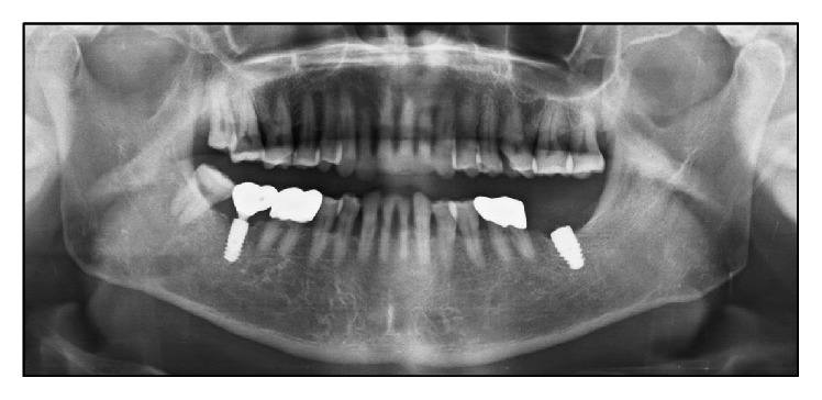
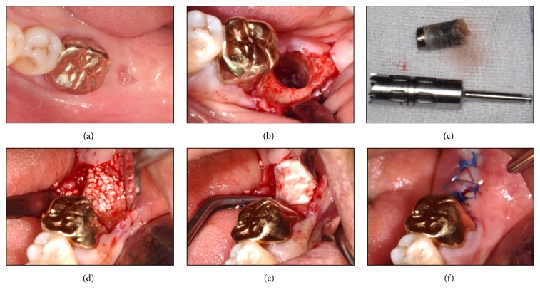
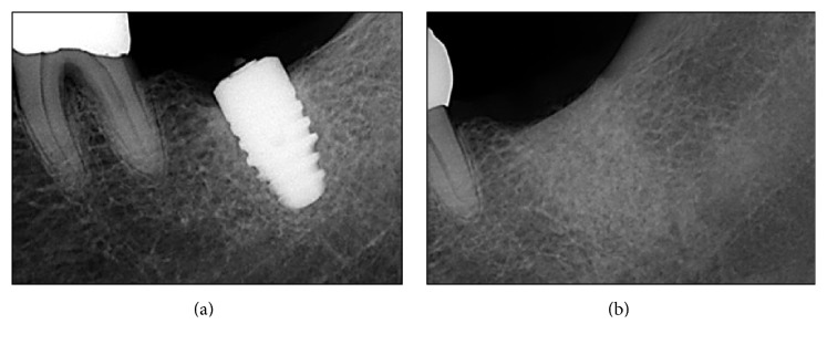
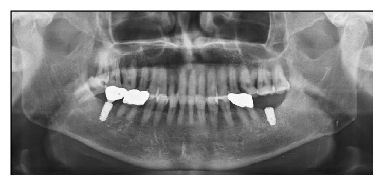
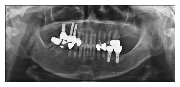
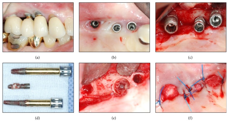
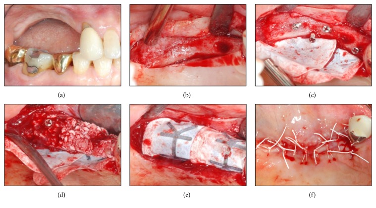
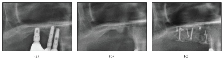
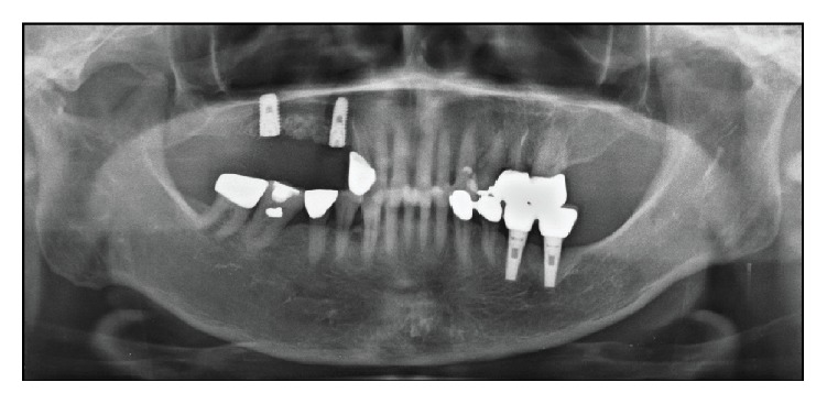
Dental implant has been restoring the function and esthetics lost from missing tooth. However, biomechanical implant complications are the major cause of failing implants. Therefore, implant removal is one of the indispensable dental treatments. The 70-year-old male and 66-year-old female who had discomfort on posterior implants region came to Department of Periodontology. Conventional method using trephine bur and the new, nontraumatic method using a fixture removal kit were used for implant removal, respectively. Two different methods are commonly used for implant removal. Each has advantages and disadvantages; thus, the applied surgical method must consider a patient's intraoral condition, posttreatment plan, and the level of surgeon's skill and experience. In conclusion, strategically executing the most optimal implant removal method plays a pivotal role in maximizing the success rate of implant reinstallation that follows afterwards.


Figures

Figure 1
Figure 1
Preoperative radiographic view (panoramic X-ray image). Implant of #37 area shows the broken abutment screw off the implant fixture inside.
Figure 2
Figure 2
Conventional removal method applied on #37 region. (a) Preoperative state showing a gingival defect. (b) Implant removal using a trephine bur (external diameter: 6 mm, internal diameter: 5 mm; Biomet, Warsaw, IN, USA), elevator (Hu-Friedy, Chicago, IL, USA), and root forceps (Hu-Friedy, Chicago, IL, USA). (c) The implant and the surrounding alveolar bone removal. ((d) and (e)) Immediate bone grafting [Osteon II 0.5 g (particle size 0.5~1.0 mm) and collagen membrane 15 × 20 mm; Genoss, Suwon, Korea] on the implant removal socket. (f) Suture for a primary wound closure.
Figure 3
Figure 3
Postoperative radiographic view (periapical X-ray image). (a) Implant fixture with a fractured abutment screw. (b) Implant removal site filled with a bone grafting material.
Figure 4
Figure 4
Postoperative radiographic view (panoramic X-ray image). Implant reinstallation on #37 region four months after the implant removal.
Figure 5
Figure 5
Preoperative radiographic view (panoramic X-ray image). Implants of right maxilla area show a severe alveolar bone loss lesion.
Figure 6
Figure 6
Advanced and mixed removal methods applied on #14, #15, and #16 regions. (a) Supragingival eruption state of an implant fixture. (b) Implant prosthodontics removal state showing a lack of keratinized gingiva and peri-implant tissue impairment. (c) Exposure of upper portion of the implant fixture and severe surrounding alveolar bone loss. (d) Implant removal using the Neo FR Kit (NeoBiotech, Seoul, Korea) on #14 and #16 regions and using the trephine bur (external diameter: 5 mm, internal diameter: 4 mm) on #15 region and the surrounding alveolar bone. (e) Implant removal socket showing a perforation of the sinus inferior wall and maintenance of the internal schneiderian membrane. (f) Tension-free suture for a natural healing progress.
Figure 7
Figure 7
Vertical augmentation of alveolar ridge applied on the implant removal site. (a) Severe vertical resorption state four months after the implant removal. (b) Narrow alveolar ridge. (c) Insertion of a tenting screw (Dentium, Seoul, Korea) and application of a GORE-TEX membrane TR6Y (W. L. Gore & Associates Inc., Flagstaff, Arizona, USA). ((d) and (e)) Bone grafting using a Bio-Oss 1.0 g (Geistlich Pharma AG, Wolhusen, Switzerland), and GORE-TEX membrane covering the site and supported by the tenting screw. (f) Suture for a primary wound closure.
Figure 8
Figure 8
Postoperative radiographic view (periapical X-ray image). (a) Implant fixture with severe surrounding alveolar bone loss. (b) Implant removal site. (c) Vertical augmentation of an alveolar ridge.
Figure 9
Figure 9
Postoperative radiographic view (panoramic X-ray image). Implant reinstallation on #14 and #16 regions four months after the implant removal.

References

    1. Pjetursson B. E., Tan K., Lang N. P., Brägger U., Egger M., Zwahlen M. A systematic review of the survival and complication rates of fixed partial dentures (FPDs) after an observation period of at least 5 years IV. Cantilever or extension FPDs. Clinical Oral Implants Research. 2004;15(6):667–676. doi: 10.1111/j.1600-0501.2004.01120.x. - DOI - PubMed
    1. Buser D., Ingimarsson S., Dula K., Lussi A., Hirt H. P., Belser U. C. Long-Term Stability of Osseointegrated Implants in Augmented Bone: A 5-Year Prospective Study in Partially Edentulous Patients. International Journal of Periodontics and Restorative Dentistry. 2002;22(2):108–117. - PubMed
    1. Carlson B., Carleson G. E. Prosthodontic complications in osseointegrated dental implant treatment. International Journal of Oral and Maxillofacial Implants. 1994;9(1):90–94. - PubMed
    1. Jung R. E., Zembic A., Pjetursson B. E., Zwahlen M., Thoma D. S. Systematic review of the survival rate and the incidence of biological, technical, and aesthetic complications of single crowns on implants reported in longitudinal studies with a mean follow-up of 5 years. Clinical Oral Implants Research. 2012;23(6):2–21. doi: 10.1111/j.1600-0501.2012.02547.x. - DOI - PubMed
    1. Jung R. E., Pjetursson B. E., Glauser R., Zembic A., Zwahlen M., Lang N. P. A systematic review of the 5-year survival and complication rates of implant-supported single crowns. Clinical Oral Implants Research. 2008;19(2):119–130. doi: 10.1111/j.1600-0501.2007.01453.x. - DOI - PubMed

Publication types

LinkOut - more resources