Review
. 2017 Aug 30;34(9):1141-1172.
doi: 10.1039/c7np00034k.

Cytochromes P450 for natural product biosynthesis in Streptomyces: sequence, structure, and function

Affiliations
Review

Cytochromes P450 for natural product biosynthesis in Streptomyces: sequence, structure, and function

Jeffrey D Rudolf et al. Nat Prod Rep. .

Abstract

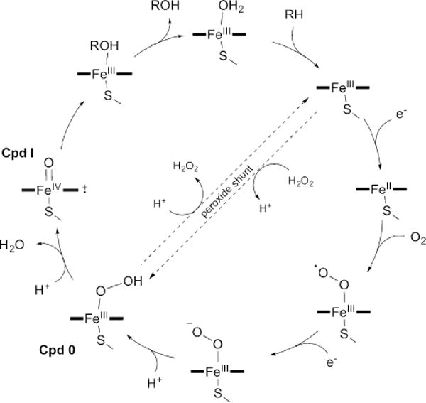
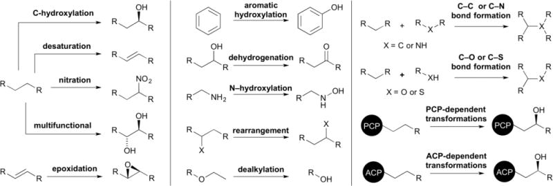
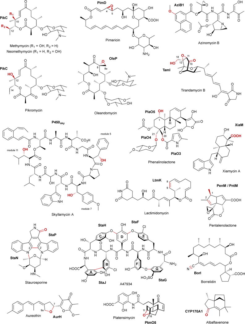
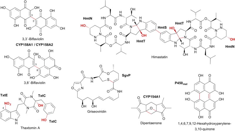
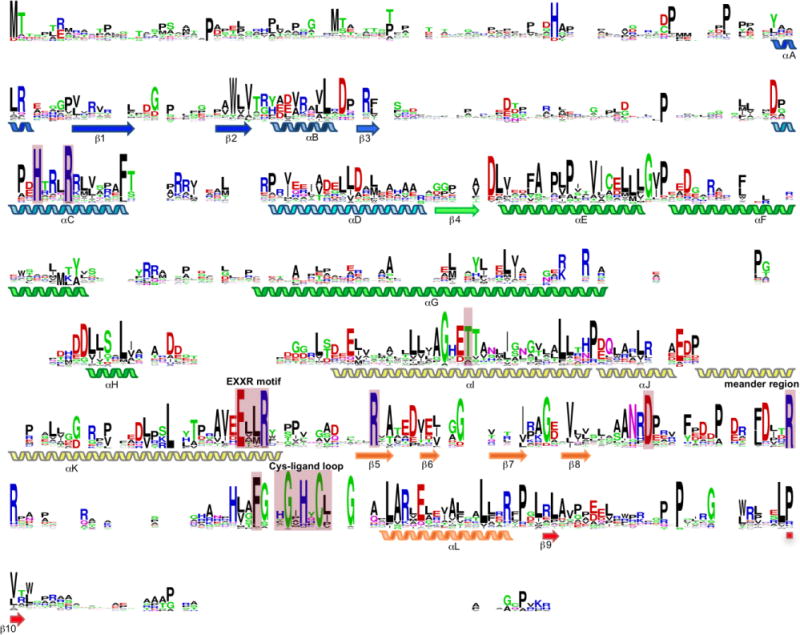
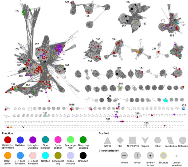
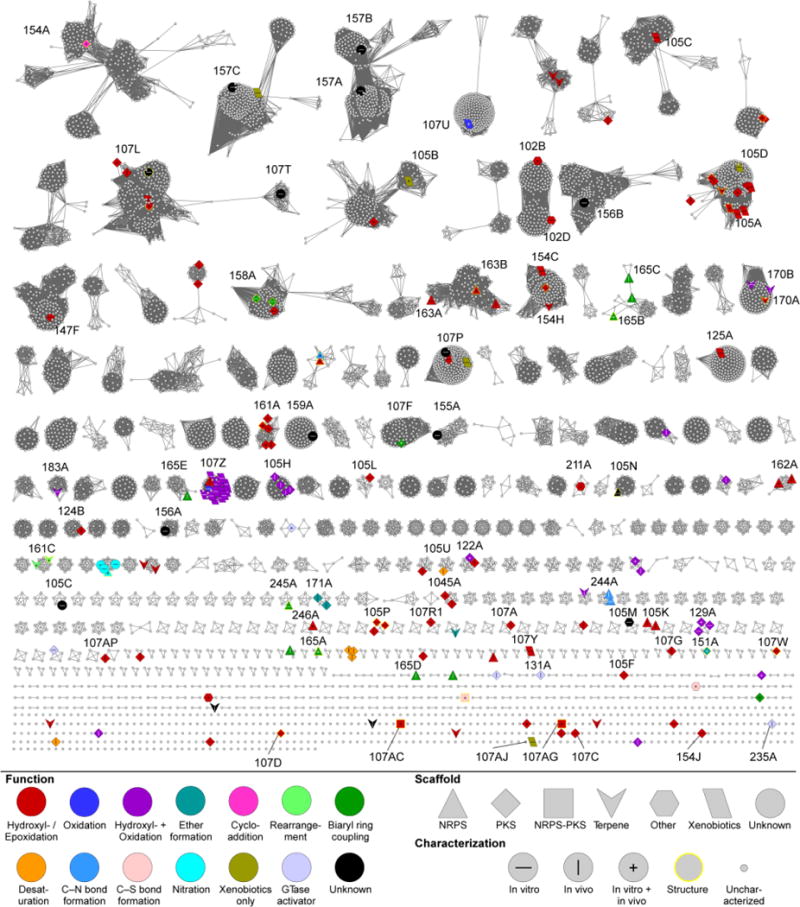
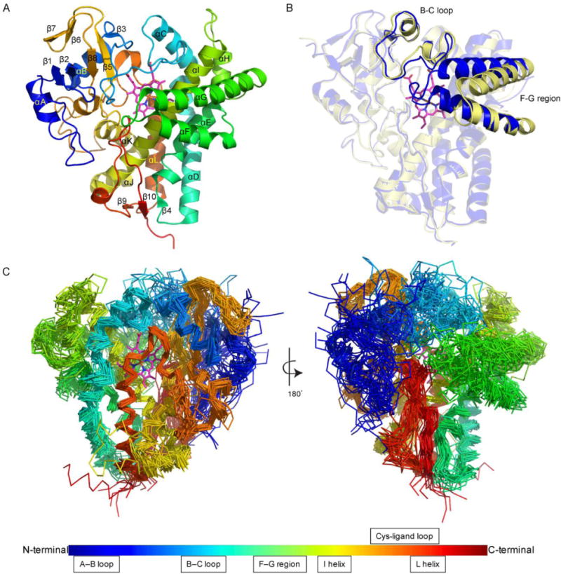
Covering: up to January 2017Cytochrome P450 enzymes (P450s) are some of the most exquisite and versatile biocatalysts found in nature. In addition to their well-known roles in steroid biosynthesis and drug metabolism in humans, P450s are key players in natural product biosynthetic pathways. Natural products, the most chemically and structurally diverse small molecules known, require an extensive collection of P450s to accept and functionalize their unique scaffolds. In this review, we survey the current catalytic landscape of P450s within the Streptomyces genus, one of the most prolific producers of natural products, and comprehensively summarize the functionally characterized P450s from Streptomyces. A sequence similarity network of >8500 P450s revealed insights into the sequence-function relationships of these oxygen-dependent metalloenzymes. Although only ∼2.4% and <0.4% of streptomycete P450s have been functionally and structurally characterized, respectively, the study of streptomycete P450s involved in the biosynthesis of natural products has revealed their diverse roles in nature, expanded their catalytic repertoire, created structural and mechanistic paradigms, and exposed their potential for biomedical and biotechnological applications. Continued study of these remarkable enzymes will undoubtedly expose their true complement of chemical and biological capabilities.


Conflict of interest statement

Conflicts of Interest

There are no conflicts of interest to declare.

Figures

Fig. 1
Fig. 1
The cytochrome P450 catalytic cycle. The cycle, shown here depicting hydroxylation of the substrate RH to yield the product ROH, is described in the text. The peroxide shunt pathway can directly form Cpd 0 from the substrate-bound high-spin FeIII state using H2O2.
Fig. 2
Fig. 2
The functional diversity of P450s in Streptomyces. P450s catalyze a wide variety of functionalizations (selected examples are shown) in natural product biosynthetic pathways and in xenobiotic degradation.
Fig. 3
Fig. 3
Selected natural products, P450s, and the biosynthetic transformations they catalyze, as discussed in the text. Functional groups and bonds colored in red are catalyzed by the P450s labeled in bold.
Fig. 3
Fig. 3
Selected natural products, P450s, and the biosynthetic transformations they catalyze, as discussed in the text. Functional groups and bonds colored in red are catalyzed by the P450s labeled in bold.
Fig. 4
Fig. 4
Sequence alignment of the 184 functionally characterized P450s from Streptomyces (minus CYP102D1, for simplicity, given its extended length). The heme-binding Cys, EXXR motif in the K-helix, and Thr in the I-helix are highly conserved. In addition, there are other highly conserved motifs and residues, both within and outside of the active site, in Streptomyces P450s. Residues discussed in the text are highlighted by pink boxes. Positions with no residues represent gaps in the alignment due to sequence length differences.
Fig. 5
Fig. 5
CYP family SSN of Streptomyces P450s. The SSN is shown at a BLAST E value cutoff = 10−85 (median 45% identity over 400 residues). Larger nodes are functionally characterized P450s with node labels describing how it was characterized. Colors and shapes of nodes represent P450 function and substrate type (type of natural product scaffold). See inset legend for details. CYP families of functionally characterized P450s are labeled.
Fig. 6
Fig. 6
CYP subfamily SSN of Streptomyces P450s. The SSN is shown at a BLAST E value cutoff = 10−124 (median 58% identity over 400 residues). Larger nodes are functionally characterized P450s with node labels describing how it was characterized. Colors and shapes of nodes represent P450 function and substrate type (type of natural product scaffold). See inset legend for details. CYP subfamilies of functionally characterized P450s are labeled.
Fig. 7
Fig. 7
Common structural aspects of P450s from Streptomyces. (A) The overall structure of P450s (exemplified by PDB ID: 3ABA). (B) The open and closed states of the P450 active site are facilitated by conformational changes in the B–C loop and the F–G region [PDB IDs: 1SE6 (open) and 2D09 (closed)]. (C) Structural superposition of the 29 structurally characterized P450s from Streptomyces highlighting the regions of high, moderate, and low structural conservativity.
Fig. 8
Fig. 8
Selected variations in the structures of P450s from Streptomyces. (A) Typical heme orientations and inversed orientations in four different P450s (PDB IDs: 1ODO, 5EX8, 4MM0, 5IT1). (B) The crystal structure of P450sky in complex with an inhibitor-tethered PCP domain (PDB ID: 4PXH). The inset shows a zoomed-in look at the interface of P450sky and the PCP domain highlighting the hydrophobic and electrostatic interactions. The unusual C-terminal M-helix is labeled. (C) Structural superposition of StaF (PDB ID: 5EX8) and StaH (PDB ID: 5EX6) with OxyB in complex with the X-domain (PDB ID: 4TX3). The inset shows the two Asp residues from the conserved PRDD motif, which are proposed to be involved in recruitment by the X-domain via interaction with two Arg residues from the X-domain. (D) The crystal structure of CYP170A1 (PDB ID: 3EL3). The B–C loop and F–G region, colored in yellow and green, respectively, form the P450 active site. The four helices, colored in red, form the terpene synthase active site.
Fig. 9
Fig. 9
Structure-based mechanistic studies of P450s from Streptomyces. (A) Epoxidation by PimD via the hydroperoxyferric intermediate, Cpd 0 (PDB ID: 2XBK). (B) Substrate-assisted biaryl ring coupling by CYP158A1 and CYP158A2 (PDB IDs: 2NZ5 and 2D09). The two different binding modes of the two biflaviolin substrates are shown in orange and green. (C) Intramolecular biaryl ring coupling by StaP (PDB IDs: 2Z3U). Wat644 and His250 are shown in the active site; Wat789 is liberated during the formation of Cpd I (Wang 2009). (D) Oxidative rearrangement by PntM via a carbocation intermediate (PDB IDs: 5L1O). Blue dots in each figure represent water molecules; yellow spheres depict steric hindrance.
Fig. 10
Fig. 10
Structure-based engineering of P450s in Streptomyces. (A) Functional switch, from hydroxylation and ether formation to hydroxylation and oxidation to carboxylic acid, of AurH via site-directed mutagenesis. (B) Nitration regioselectivity switch, from C-4 to C-5, of TxtE via site-directed mutagenesis. (C) Substrate engineering of PikC shifting the natural abundance of regioisomers. The ratios representing the C-10:C-12 hydroxylated products changes from 1:1 with the natural desosamine anchoring group to 1:4 or >20:1 depending on the synthetic anchoring variant used.

References

    1. Guengerich FP. In: Cytochrome P450: Structure, Mechanism, and Biochemistry. 4th. Ortiz de Montellano PR, editor. Springer International Publishing; Switzerland: 2015. pp. 523–785.
    1. Auchus RJ, Miller WL. In: Cytochrome P450: Structure, Mechanism, and Biochemistry. 4th. Ortiz de Montellano PR, editor. Springer International Publishing; Switzerland: 2015. pp. 851–879.
    1. Podust LM, Sherman DH. Nat Prod Rep. 2012;29:1251–1266. - PMC - PubMed
    1. McLean KJ, Leys D, Munro AW. In: Cytochrome P450: Structure, Mechanism, and Biochemistry. 4th. Ortiz de Montellano PR, editor. Springer International Publishing; Switzerland: 2015. pp. 261–407.
    1. Schuler MA. In: Cytochrome P450: Structure, Mechanism, and Biochemistry. 4th. Ortiz de Montellano PR, editor. Springer International Publishing; Switzerland: 2015. pp. 409–449.

Publication types

MeSH terms

Substances