Case Reports
. 2017 Dec;23(6):614-619.
doi: 10.1177/1591019917722514. Epub 2017 Jul 31.

Pipeline embolization device retraction and foreshortening after internal carotid artery blister aneurysm treatment

Affiliations
Case Reports

Pipeline embolization device retraction and foreshortening after internal carotid artery blister aneurysm treatment

Jeremy J Heit et al. Interv Neuroradiol. 2017 Dec.

Abstract

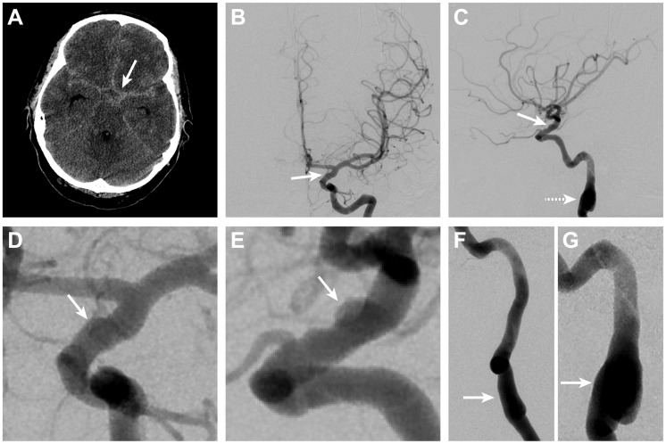
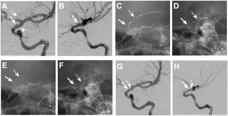
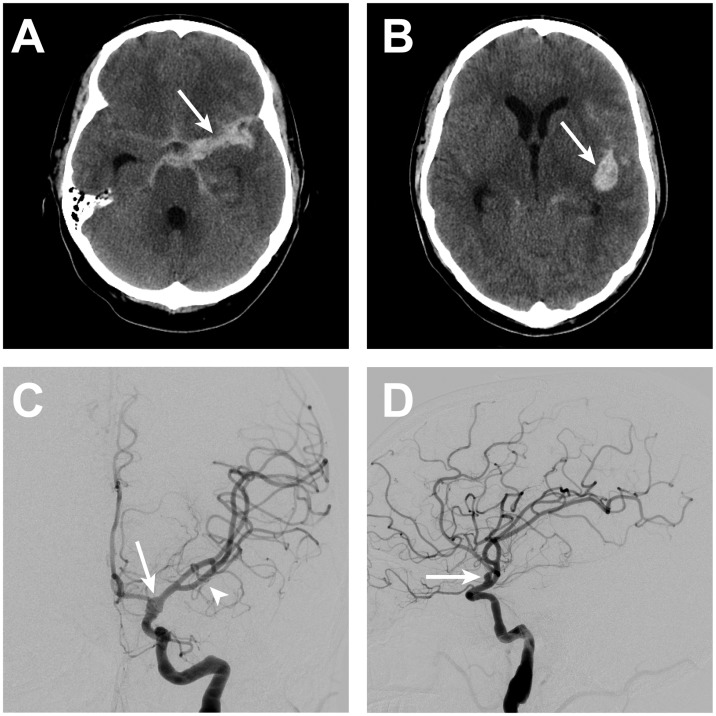
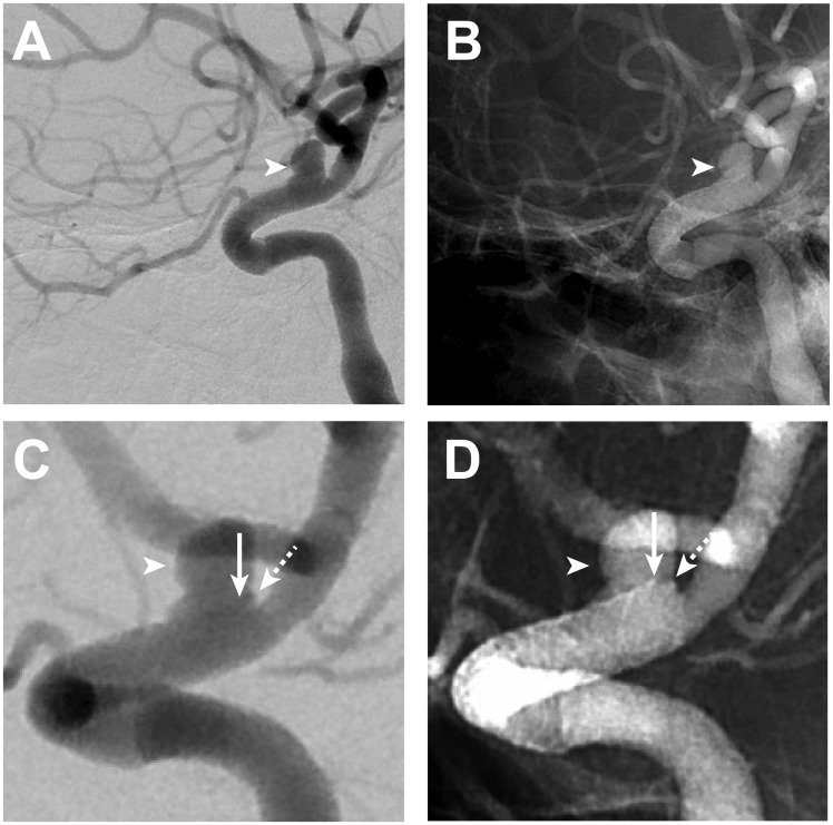
Background Subarachnoid hemorrhage (SAH) secondary to rupture of a blister aneurysm (BA) results in high morbidity and mortality. Endovascular treatment with the pipeline embolization device (PED) has been described as a new treatment strategy for these lesions. We present the first reported case of PED retraction and foreshortening after treatment of a ruptured internal carotid artery (ICA) BA. Case description A middle-aged patient presented with SAH secondary to ICA BA rupture. The patient was treated with telescoping PED placement across the BA. After 5 days from treatment, the patient developed a new SAH due to re-rupture of the BA. Digital subtraction angiography revealed an increase in caliber of the supraclinoid ICA with associated retraction and foreshortening of the PED that resulted in aneurysm uncovering and growth. Conclusions PED should be oversized during ruptured BA treatment to prevent device retraction and aneurysm regrowth. Frequent imaging follow up after BA treatment with PED is warranted to ensure aneurysm occlusion.

Keywords: Blister aneurysm; embolization; foreshortening; pipeline; subarachnoid hemorrhage.


Figures

Figure 1.
Figure 1.
Subarachnoid hemorrhage due to rupture of a left ICA blister aneurysm. Non-contrast head CT shows diffuse SAH in the basal cisterns (arrow, A). Left ICA DSA in the anteroposterior (B) and lateral projections (C) shows irregularity with an associated pseudoaneurysm (arrows, B and C) on the dorsal wall of the left ICA, which is consistent with a BA. Magnified DSA views in the anteroposterior (D) and lateral (E) projections better demonstrate the BA (arrows, D and E). Irregularity that is consistent with prior dissection (arrows, F and G) was also present in the right (F) and left (G) cervical internal carotid arteries on DSA, which suggests the presence of an underlying connective tissue disorder. BA: blister aneurysm; CT: computed tomography; DSA: digital subtraction angiography; ICA: internal carotid artery; SAH: subarachnoid hemorrhage
Figure 2.
Figure 2.
ICA blister aneurysm treatment with telescoping PED. Left ICA DSA images from oblique anteroposterior (A) and lateral (B) projections show the BA (arrows, A and B) arising from the dorsal wall of the left ICA. The distal aspect of the BA terminates at the aneurysm edge just proximal to the origin (arrowhead, A) of the left anterior choroidal artery (dashed arrow, A). Fluoroscopic images in oblique anteroposterior (C) and lateral (D) projections show the first PED (arrows, C and D) deployed across the BA while a second PED (dashed arrows, C and D) is being deployed distal to the termination of the first PED. The second PED (dashed arrows, E and F) terminates at the left ICA apex, which is 6 mm distal to the distal aspect of the first PED (arrow, E and F) after deployment. DSA following telescoping PED placement demonstrates persistent filling of the aneurysm (arrows, G and H); the distal most aspect of the telescoped PEDs is indicated by the dashed arrow (G, H). BA: blister aneurysm; DSA: digital subtraction angiography; ICA: internal carotid artery; PED: pipeline embolization device
Figure 3.
Figure 3.
ICA blister aneurysm re-rupture. After 5 days from BA treatment with telescoping PED, the patient suffered aneurysm re-rupture that resulted in new SAH within the basal cisterns (arrow, A) and the left Sylvian fissure (arrow, B). Left ICA DSA in the anteroposterior (C) and lateral (D) projections demonstrated an increase in size of the left ICA blister aneurysm/pseudoaneurysm (arrows, C and D). Mass effect on the left middle cerebral artery vessels from the Sylvian fissure hemorrhage is apparent on the DSA with superior and medial displacement of these vessels (arrowhead, C). BA: blister aneurysm; DSA: digital subtraction angiography; ICA: internal carotid artery; PED: pipeline embolization device; SAH: subarachnoid hemorrhage
Figure 4.
Figure 4.
ICA blister aneurysm growth after re-rupture. All left ICA DSA images (A–D) are in an oblique lateral projection. Left ICA DSA performed after BA re-rupture (A–D) shows an interval increase in size of the left ICA blister aneurysm/pseudoaneurysm (arrowheads, A–D) compared with the left ICA DSA performed immediately after telescoping PED placement (see Figure 2). Magnified subtracted (C) and unsubtracted images (D) on the second DSA better demonstrate PED retraction and foreshortening by approximately 7 mm with the superior aspect of the distal PED construct. The PED is slightly obliqued on these views, such that the distal end of the PED projects with a portion of the distal edge of the PED located 1 mm proximal to the distal edge of the BA (arrows, C and D) and the other portion of the distal PED to be aligned with the distal end of the BA (dashed arrows, C and D). BA: blister aneurysm; DSA: digital subtraction angiography; ICA: internal carotid artery; PED: pipeline embolization device
Figure 5.
Figure 5.
Endovascular coil sacrifice of the left ICA for recurrent blister aneurysm treatment. Endovascular occlusion of the supraclinoid left ICA within and just distal to the previously placed telescoping PEDs was performed following re-rupture of the left ICA BA. The coil mass (arrows, A and B) are present on anteroposterior and lateral radiographic images after occlusion. Right ICA DSA (C, D) performed after coil sacrifice of the left ICA shows robust filling of the left anterior circulation across a patent anterior communicating artery complex. There is filling of the distal left ICA to the margin of the coil mass (arrows, C and D) following right ICA DSA, but there no retrograde filling of the left ICA BA. BA: blister aneurysm; DSA: digital subtraction angiography; ICA: internal carotid artery; PED: pipeline embolization device

Similar articles

Cited by

References

    1. Szmuda T, Sloniewski P, Waszak PM, et al. Towards a new treatment paradigm for ruptured blood blister-like aneurysms of the internal carotid artery? A rapid systematic review. J Neurointerv Surg 2016; 8: 488–94. - PubMed
    1. Rouchaud A, Brinjikji W, Cloft HJ, et al. Endovascular treatment of ruptured blister-like aneurysms: A systematic review and meta-analysis with focus on deconstructive versus reconstructive and flow-diverter treatments. Am J Neuroradiol 2015; 36: 2331–2339. - PMC - PubMed
    1. Nerva JD, Morton RP, Levitt MR, et al. Pipeline embolization device as primary treatment for blister aneurysms and iatrogenic pseudoaneurysms of the internal carotid artery. J Neurointerv Surg 2015; 7: 210–216. - PubMed
    1. Linfante I, Mayich M, Sonig A, et al. Flow diversion with pipeline embolic device as treatment of subarachnoid hemorrhage secondary to blister aneurysms: Dual-center experience and review of the literature. J Neurointerv Surg 2017; 9: 29–33. - PubMed
    1. Hu YC, Chugh C, Mehta H, et al. Early angiographic occlusion of ruptured blister aneurysms of the internal carotid artery using the pipeline embolization device as a primary treatment option. J Neurointerv Surg 2014; 6: 740–743. - PubMed

Publication types

MeSH terms

LinkOut - more resources