. 2017 Aug;98(8):2069-2078.
doi: 10.1099/jgv.0.000869. Epub 2017 Jul 31.

Tree shrew, a potential animal model for hepatitis C, supports the infection and replication of HCV in vitro and in vivo

Affiliations

Tree shrew, a potential animal model for hepatitis C, supports the infection and replication of HCV in vitro and in vivo

Yue Feng et al. J Gen Virol. 2017 Aug.

Abstract

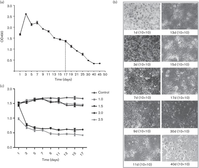
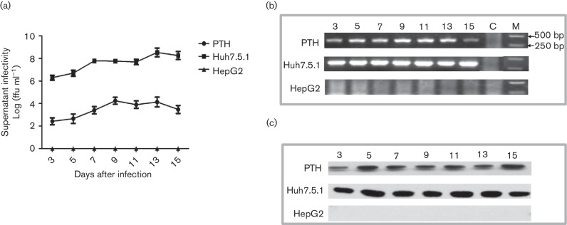
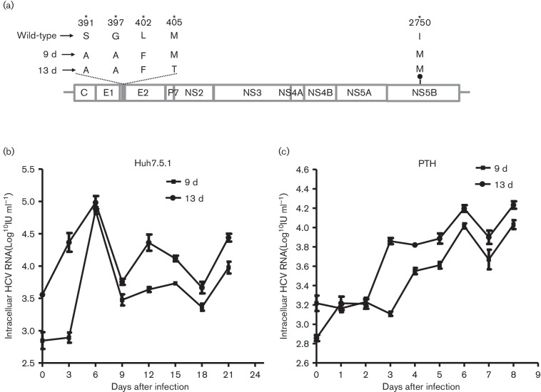
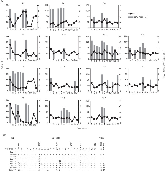
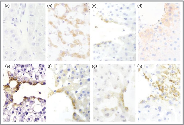
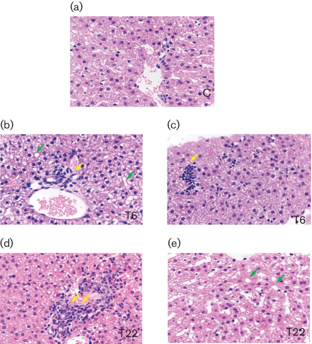
The tree shrew (Tupaia belangeri chinensis), a small animal widely distributed in Southeast Asia and southwest China, has the potential to be developed as an animal model for hepatitis C. To determine the susceptibility of the tree shrew to hepatitis C virus (HCV) infection in vitro and in vivo, a well-established HCV, produced from the J6/JFH1-Huh7.5.1 culture system, was used to infect cultured primary tupaia hepatocytes (PTHs) and tree shrews. The in vitro results showed that HCV genomic RNA and HCV-specific nonstructural protein 5A (NS5A) could be detected in the PTH cell culture from days 3-15 post-infection, although the viral load was lower than that observed in Huh7.5.1 cell culture. The occurrence of five sense mutations [S391A, G397A, L402F and M405T in the hypervariable region 1 (HVR1) of envelope glycoprotein 2 and I2750M in NS5B] suggested that HCV undergoes genetic evolution during culture. Fourteen of the 30 experimental tree shrews (46.7 %) were found to be infected, although the HCV viremia was intermittent in vivo. A positive test for HCV RNA in liver tissue provided stronger evidence for HCV infection and replication in tree shrews. The results of an immunohistochemistry assay also demonstrated the presence of four HCV-specific proteins (Core, E2, NS3/4 and NS5A) in the hepatocytes of infected tree shrews. The pathological changes observed in the liver tissue of infected tree shrews could be considered to be representative symptoms of mild hepatitis. These results revealed that the tree shrew can be used as an animal model supporting the infection and replication of HCV in vitro and in vivo.


Figures

Fig. 1.
Fig. 1.
Culture and growth of primary tupaia hepatocytes (PTHs) and effects of HCV infection. Using an MTT assay, the viability of PTHs was determined over a period of more than 30 days, with obvious cell proliferation for 3–9 days in culture (a). The activity and purity of the obtained cells and the cell growth were observed under a microscope (b). HCV infection at an m.o.i. of 2.0 and 2.5 significantly reduced cell viability, while at an m.o.i. of 1.0 and 1.5 there was little effect on the viability of PTH cells (c).
Fig. 2.
Fig. 2.
HCV infection and replication in primary tupaia hepatocytes (PTHs). HCVcc was able to infect PTHs, but showed a lower level of replication compared with that in Huh7.5.1 cells (a). HCV RNA (b) and a specific protein (c) were positively detected in the culture supernatants and cell lysates of infected PTHs using RT–nPCR and Western blot, respectively.
Fig. 3.
Fig. 3.
HCV adaptive mutations in PTHs and changes in HCV infectivity. Five sense mutations (S391A, G397A, L402F and M405T in the hypervariable region 1 (HVR1) of E2, and I2750M in NS5B) were revealed in the virus isolated on days 9 and 13 post-infection (a). Then, the media containing mutant viruses from days 9 and 13 were transferred to naïve Huh7.5.1 and PTH cells, and the same mutations were found when infection with the passaged virus was established in these two cell lines (b, c).
Fig. 4.
Fig. 4.
ALT levels and HCV viral load in tree shrews following inoculation with HCV. A total of 14 animals (46.7 %) developed intermittent HCV viremia, as determined by RT–qPCR in the 24 weeks post-inoculation. The ALT levels in these 14 infected animals did not show any obvious changes.
Fig. 5.
Fig. 5.
Immunohistochemical detection of HCV-specific proteins using a anti-substance P (SP) method. In the liver tissue of a representative animal (T22), which was infected with Huh7.5.1-produced HCV, E2 (e), core (f), NS5A (g) and NS3/4 (h) proteins were detected in the cytoplasm of the hepatocytes surrounding the central vein of the liver lobule (DAB dark yellow granular cytoplasmic staining, 400×). The same positive NS5A staining pattern is also obvious in the tissue of a hepatitis C patient (b) and a tree shrew infected with a clinical HCV strain (c). The red APE colour (d) also shows the presence of the NS5A protein in the hepatocytes of T22. By comparison, no NS5A staining was observed in the liver tissue of the negative control tree shrew (a).
Fig. 6.
Fig. 6.
Micrographs of liver specimens stained with H and E (40×). Liver tissue was collected from HCV-infected tree shrews, with T6 and T22 as representative animals, 24 weeks post-inoculation. Liver specimens from uninfected animals (control group), matched to infected animals, were also obtained. The HCV-infected tupaia livers harboured infiltrating lymphocytes (yellow arrowheads) and showed microvesicular fat accumulation (green arrowheads).

Similar articles

Cited by

References

    1. Bandiera S, Billie Bian C, Hoshida Y, Baumert TF, Zeisel MB. Chronic hepatitis C virus infection and pathogenesis of hepatocellular carcinoma. Curr Opin Virol. 2016;20:99–105. doi: 10.1016/j.coviro.2016.09.010. - DOI - PMC - PubMed
    1. Esposito I, Trinks J, Soriano V. Hepatitis C virus resistance to the new direct-acting antivirals. Expert Opin Drug Metab Toxicol. 2016;12:1197–1209. doi: 10.1080/17425255.2016.1209484. - DOI - PubMed
    1. Vercauteren K, de Jong YP, Meuleman P. Animal models for the study of HCV. Curr Opin Virol. 2015;13:67–74. doi: 10.1016/j.coviro.2015.04.009. - DOI - PMC - PubMed
    1. Bility MT, Zhang L, Washburn ML, Curtis TA, Kovalev GI, et al. Generation of a humanized mouse model with both human immune system and liver cells to model hepatitis C virus infection and liver immunopathogenesis. Nat Protoc. 2012;7:1608–1617. doi: 10.1038/nprot.2012.083. - DOI - PMC - PubMed
    1. Douglas DN, Kneteman NM. Generation of improved mouse models for the study of hepatitis C virus. Eur J Pharmacol. 2015;759:313–325. doi: 10.1016/j.ejphar.2015.03.022. - DOI - PubMed

Substances

LinkOut - more resources