. 2017 Jul 29;8(3):31.
doi: 10.3390/jfb8030031.

Effect of a Particulate and a Putty-Like Tricalcium Phosphate-Based Bone-grafting Material on Bone Formation, Volume Stability and Osteogenic Marker Expression after Bilateral Sinus Floor Augmentation in Humans

Affiliations

Effect of a Particulate and a Putty-Like Tricalcium Phosphate-Based Bone-grafting Material on Bone Formation, Volume Stability and Osteogenic Marker Expression after Bilateral Sinus Floor Augmentation in Humans

Christine Knabe et al. J Funct Biomater. .

Abstract

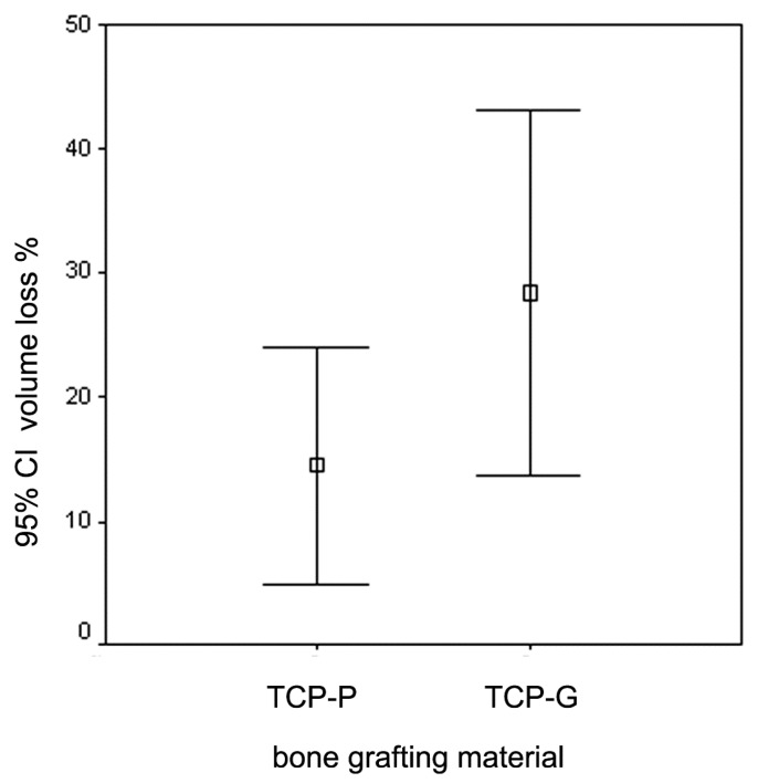
This study examines the effect of a hyaluronic acid (HyAc) containing tricalcium phosphate putty scaffold material (TCP-P) and of a particulate tricalcium phosphate (TCP-G) graft on bone formation, volume stability and osteogenic marker expression in biopsies sampled 6 months after bilateral sinus floor augmentation (SFA) in 7 patients applying a split-mouth design. 10% autogenous bone chips were added to the grafting material during surgery. The grain size of the TCP granules was 700 to 1400 µm for TCP-G and 125 to 250 µm and 500 to 700 µm (ratio 1:1) for TCP-P. Biopsies were processed for immunohistochemical analysis of resin-embedded sections. Sections were stained for collagen type I (Col I), alkaline phosphatase (ALP), osteocalcin (OC) and bone sialoprotein (BSP). Furthermore, the bone area and biomaterial area fraction were determined histomorphometrically. Cone-beam CT data recorded after SFA and 6 months later were used for calculating the graft volume at these two time points. TCP-P displayed more advantageous surgical handling properties and a significantly greater bone area fraction and smaller biomaterial area fraction. This was accompanied by significantly greater expression of Col I and BSP and in osteoblasts and osteoid and a less pronounced reduction in grafting volume with TCP-P. SFA using both types of materials resulted in formation of sufficient bone volume for facilitating stable dental implant placement with all dental implants having been in function without any complications for 6 years. Since TCP-P displayed superior surgical handling properties and greater bone formation than TCP-G, without the HyAc hydrogel matrix having any adverse effect on bone formation or graft volume stability, TCP-P can be regarded as excellent grafting material for SFA in a clinical setting. The greater bone formation observed with TCP-P may be related to the difference in grain size of the TCP granules and/or the addition of the HyAc.

Keywords: bioactive ceramics; bone formation; bone-grafting materials; hard tissue histology; immunohistochemical analysis; osteogenesis; osteogenic markers; sinus floor augmentation; split-mouth design; tricalcium phosphate putty scaffold.


Conflict of interest statement

The authors declare no conflict of interest.

Figures

Figure 1
Figure 1
Two-dimensional radiographic image generated from the secondary cone beam CT acquired directly after SFA showing of a cross section through the augmented sinus floor (red) as well as the osseous anatomical structures of the native sinus floor (blue). This image was generated by merging the data sets of the primary and secondary cone-beam CTs. Sagittal view of the, left-sided maxillary sinus, patient 5.
Figure 2
Figure 2
Two-dimensional radiographic image generated from the tertiary cone-beam CT acquired at implant placement 6 months after SFA showing of a cross section through grafted area after the 6 months healing period (green) as well as the osseous anatomical structures of the native sinus floor (blue). This image was generated by merging the data sets of the primary and tertiary cone-beam CTs in combination with superimposing the image of the secondary cone beam CT (light grey). Sagittal view of the left-sided maxillary sinus, patient 5.
Figure 3
Figure 3
Decrease (%) in grafting volume observed 6 months after sinus floor augmentation with TCP-putty and TCP granules in 7 individual patients.
Figure 4
Figure 4
Graph depicting the mean values and 95% confidence interval (CI) for the decrease in grafting volume 6 months after SFA using CEROS®-TCP-putty (TCP-P) and CEROS®-TCP-granules (TCP-G).
Figure 5
Figure 5
Histomicrograph of resin embedded biopsy stained immunohistochemically for osteocalcin after deacrylation. The biopsy was sampled 6 months after augmentation of the sinus floor with TCP-G (B = bone, FM = fibrous matrix of the osteogenic mesenchym, TCP-G—residual TCP particles displaying a scalloped morphology). Undecalcified sawed section counterstained with hematoxylin. Bar = 2000 µm.
Figure 6
Figure 6
Histomicrograph of resin embedded biopsy stained immunohistochemically for osteocalcin after deacrylation. The biopsy was sampled 6 months after SFA with TCP-P (B = bone, FM = fibrous matrix, TCP-P = residual TCP particles of the putty scaffold material. The smaller grain size of these particles compared to the TCP-G material is evident. Undecalcified sawed section counterstained with hematoxylin. Bar = 2000 µm.
Figure 7
Figure 7
Histomicrographs of resin embedded biopsies sampled 6 months after SFA with TCP-P or TCP-G stained immunohistochemically for the osteogenic markers bone sialoprotein (a,b), osteocalcin (c,d), type I collagen (e,f), alkaline phosphatase (g,h) after deacrylation: (a) Immunodetection of bone sialoprotein in sawed section of biopsy sampled 6 months after SFA floor with TCP-P. Intense staining of osteoblasts, which have migrated into the degrading TCP-P particles is visible (white arrows), which exhibit excellent bone (B)-particle contact, i.e., bone-bonding behavior. Furthermore, bone formation within the degrading particles is visible (black arrow) as well as strong staining of the mineralizing but not yet fully mineralized bone matrix (black arrowhead) in contact with the TCP-P particles; (b) Immunodetection of bone sialoprotein in sawed section of biopsy sampled 6 months after SFA with TCP-G. Intense staining of osteoblasts, which have migrated into the degrading TCP-G particles is visible (white arrows), which exhibit excellent bone particle contact, i.e., bone-bonding behavior (yellow arrowheads). Furthermore, mild staining of the osteoid (black arrows) and mineralizing but not yet fully mineralized bone matrix, i.e., osteogenic mesenchym, (black arrowheads) in contact with the TCP-G particles is present; (c) Immunodetection of osteocalcin in hard tissue section of biopsy sampled 6 months after SFA floor with TCP-P. TCP-P-particles are visible, which exhibit excellent bone particle contact, (yellow arrowheads). Furthermore, strong staining of the osteoid (black arrows) and mineralizing but not yet fully mineralized bone matrix (black arrowheads) in contact with the TCP-P particles is present; (d) Immunodetection of osteocalcin in section of biopsy sampled 6 months after SFA floor with TCP-G. TCP-G-particles are present, which exhibit partial bone particle contact (yellow arrowheads). Osteoid (black arrows) and mineralizing but not yet fully mineralized bone matrix (black arrowheads) without any positive staining for osteocalcin are visible. In addition, osteoid with mild osteocalcin expression (white arrowheads) lining marrow spaces is visible. Undecalcified sawed section counterstained with hematoxylin. Bar = 200 µm; (e) Immunodetection of type I collagen in section of biopsy sampled 6 months after SFA floor with TCP-P. Intense staining of osteoblasts, which have migrated into the degrading TCP-P particles is visible (white arrows), which exhibit excellent bone-particle contact (yellow arrowheads). Furthermore, strong staining of the mineralizing but not yet fully mineralized bone matrix (black arrowheads) as well as of the fully mineralized bone matrix (green arrow) in contact with the TCP-P particles is present. Bar = 100 µm; (f) Immunodetection of type I collagen in section of biopsy sampled form TCP-G site. TCP-G particles are present, which exhibit partial bone particle contact (yellow arrowheads). Furthermore, moderate staining of the osteoid (black arrows) and mineralizing but not yet fully mineralized bone matrix (black arrowhead) in contact with the TCP-G particles is visible. This is in addition to mild staining of the mineralized matrix (green arrows); (g) Immunodetection of alkaline phosphatase in section of biopsy sampled from TCP-P site. TCP-P particles are visible, which exhibit excellent bone particle contact (yellow arrowheads). Furthermore, strong staining of the osteoblasts (white arrows) and of the mineralizing but not yet fully mineralized bone matrix (black arrowheads) in contact with the TCP-P particles is present; (h) Immunodetection of alkaline phosphatase in sawed section of biopsy sampled 6 months after SFA floor with TCP-G. TCP-G particles are present, which exhibit excellent bone particle contact (yellow arrowheads). Strong staining of the mineralizing but not yet fully mineralized bone matrix (black arrowheads) in contact with the TCP-G particles is visible. Bar = 200 µm; (i) histomicrograph of positive control section of experimental fracture site stained for TRAP activity enzymhistochemically. A TRAP-positive multinucleated osteoclast (black arrow) is visible. Bar = 20µm.
Figure 7
Figure 7
Histomicrographs of resin embedded biopsies sampled 6 months after SFA with TCP-P or TCP-G stained immunohistochemically for the osteogenic markers bone sialoprotein (a,b), osteocalcin (c,d), type I collagen (e,f), alkaline phosphatase (g,h) after deacrylation: (a) Immunodetection of bone sialoprotein in sawed section of biopsy sampled 6 months after SFA floor with TCP-P. Intense staining of osteoblasts, which have migrated into the degrading TCP-P particles is visible (white arrows), which exhibit excellent bone (B)-particle contact, i.e., bone-bonding behavior. Furthermore, bone formation within the degrading particles is visible (black arrow) as well as strong staining of the mineralizing but not yet fully mineralized bone matrix (black arrowhead) in contact with the TCP-P particles; (b) Immunodetection of bone sialoprotein in sawed section of biopsy sampled 6 months after SFA with TCP-G. Intense staining of osteoblasts, which have migrated into the degrading TCP-G particles is visible (white arrows), which exhibit excellent bone particle contact, i.e., bone-bonding behavior (yellow arrowheads). Furthermore, mild staining of the osteoid (black arrows) and mineralizing but not yet fully mineralized bone matrix, i.e., osteogenic mesenchym, (black arrowheads) in contact with the TCP-G particles is present; (c) Immunodetection of osteocalcin in hard tissue section of biopsy sampled 6 months after SFA floor with TCP-P. TCP-P-particles are visible, which exhibit excellent bone particle contact, (yellow arrowheads). Furthermore, strong staining of the osteoid (black arrows) and mineralizing but not yet fully mineralized bone matrix (black arrowheads) in contact with the TCP-P particles is present; (d) Immunodetection of osteocalcin in section of biopsy sampled 6 months after SFA floor with TCP-G. TCP-G-particles are present, which exhibit partial bone particle contact (yellow arrowheads). Osteoid (black arrows) and mineralizing but not yet fully mineralized bone matrix (black arrowheads) without any positive staining for osteocalcin are visible. In addition, osteoid with mild osteocalcin expression (white arrowheads) lining marrow spaces is visible. Undecalcified sawed section counterstained with hematoxylin. Bar = 200 µm; (e) Immunodetection of type I collagen in section of biopsy sampled 6 months after SFA floor with TCP-P. Intense staining of osteoblasts, which have migrated into the degrading TCP-P particles is visible (white arrows), which exhibit excellent bone-particle contact (yellow arrowheads). Furthermore, strong staining of the mineralizing but not yet fully mineralized bone matrix (black arrowheads) as well as of the fully mineralized bone matrix (green arrow) in contact with the TCP-P particles is present. Bar = 100 µm; (f) Immunodetection of type I collagen in section of biopsy sampled form TCP-G site. TCP-G particles are present, which exhibit partial bone particle contact (yellow arrowheads). Furthermore, moderate staining of the osteoid (black arrows) and mineralizing but not yet fully mineralized bone matrix (black arrowhead) in contact with the TCP-G particles is visible. This is in addition to mild staining of the mineralized matrix (green arrows); (g) Immunodetection of alkaline phosphatase in section of biopsy sampled from TCP-P site. TCP-P particles are visible, which exhibit excellent bone particle contact (yellow arrowheads). Furthermore, strong staining of the osteoblasts (white arrows) and of the mineralizing but not yet fully mineralized bone matrix (black arrowheads) in contact with the TCP-P particles is present; (h) Immunodetection of alkaline phosphatase in sawed section of biopsy sampled 6 months after SFA floor with TCP-G. TCP-G particles are present, which exhibit excellent bone particle contact (yellow arrowheads). Strong staining of the mineralizing but not yet fully mineralized bone matrix (black arrowheads) in contact with the TCP-G particles is visible. Bar = 200 µm; (i) histomicrograph of positive control section of experimental fracture site stained for TRAP activity enzymhistochemically. A TRAP-positive multinucleated osteoclast (black arrow) is visible. Bar = 20µm.
Figure 8
Figure 8
Histogram illustrating the results of the histomorphometric evaluation (mean values) of the area fraction of the newly formed bony trabeculae, of the biomaterial/particle area fraction and the area fraction of the bone marrow spaces in biopsies sampled bilaterally from seven patients 6 months after SFA with TCP-P and TCP-G.
Figure 9
Figure 9
Histogram depicting the results of the histomorphometric analysis (mean values ± SEM) of the area fraction of the newly formed bony trabeculae and of the particle (residual biomaterial) area fraction in biopsies obtained bilaterally from seven patients 6 months after SFA with TCP-P and TCP-G. Asterisks indicate statistical significance.
Figure 10
Figure 10
Representative panoramic radiograph showing dental implants without any marginal periimplant bone loss (yellow arrows) 6 years after implant placement in the grafted sinus floors (TCP-P, left; TCP-G, right).

Similar articles

Cited by

References

    1. Aghaloo T.L., Moy P.K. Which hard tissue augmentation techniques are the most successful in furnishing bony support for implant placement? Int. J. Oral Maxillofac. Implant. 2007;22:49–70. - PubMed
    1. Knabe C., Koch C., Rack A., Stiller M. Effect of beta-tricalcium phosphate particles with varying porosity on osteogenesis after sinus floor augmentation in humans. Biomaterials. 2008;29:2249–2258. doi: 10.1016/j.biomaterials.2008.01.026. - DOI - PubMed
    1. Pjetursson B.E., Tan W.C., Zwahlen M., Lang N.P. A systematic review of the success of sinus floor elevation and survival of implants inserted in combination with sinus floor elevation. J. Clin. Periodontol. 2008;35:216–240. doi: 10.1111/j.1600-051X.2008.01272.x. - DOI - PubMed
    1. Kalk W.W., Raghoebar G.M., Jansma J., Boering G. Morbidity from iliac crest bone harvesting. J. Oral Maxillofac. Surg. 1996;54:1424–1429. doi: 10.1016/S0278-2391(96)90257-8. - DOI - PubMed
    1. Wheeler S.L. Sinus augmentation for dental implants: The use of alloplastic materials. J. Oral Maxillofac. Surg. 1997;55:1287–1293. doi: 10.1016/S0278-2391(97)90186-5. - DOI - PubMed

LinkOut - more resources