

EMT/MET at the Crossroad of Stemness, Regeneration and Oncogenesis: The Ying-Yang Equilibrium Recapitulated in Cell Spheroids
- PMID: 28758926
- PMCID: PMC5575601
- DOI: 10.3390/cancers9080098
EMT/MET at the Crossroad of Stemness, Regeneration and Oncogenesis: The Ying-Yang Equilibrium Recapitulated in Cell Spheroids
Abstract
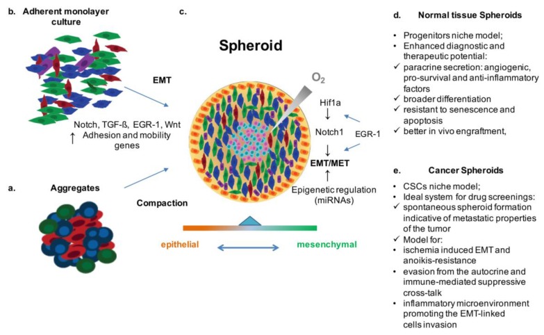
The epithelial-to-mesenchymal transition (EMT) is an essential trans-differentiation process, which plays a critical role in embryonic development, wound healing, tissue regeneration, organ fibrosis, and cancer progression. It is the fundamental mechanism by which epithelial cells lose many of their characteristics while acquiring features typical of mesenchymal cells, such as migratory capacity and invasiveness. Depending on the contest, EMT is complemented and balanced by the reverse process, the mesenchymal-to-epithelial transition (MET). In the saving economy of the living organisms, the same (Ying-Yang) tool is integrated as a physiological strategy in embryonic development, as well as in the course of reparative or disease processes, prominently fibrosis, tumor invasion and metastasis. These mechanisms and their related signaling (e.g., TGF-β and BMPs) have been effectively studied in vitro by tissue-derived cell spheroids models. These three-dimensional (3D) cell culture systems, whose phenotype has been shown to be strongly dependent on TGF-β-regulated EMT/MET processes, present the advantage of recapitulating in vitro the hypoxic in vivo micro-environment of tissue stem cell niches and their formation. These spheroids, therefore, nicely reproduce the finely regulated Ying-Yang equilibrium, which, together with other mechanisms, can be determinant in cell fate decisions in many pathophysiological scenarios, such as differentiation, fibrosis, regeneration, and oncogenesis. In this review, current progress in the knowledge of signaling pathways affecting EMT/MET and stemness regulation will be outlined by comparing data obtained from cellular spheroids systems, as ex vivo niches of stem cells derived from normal and tumoral tissues. The mechanistic correspondence in vivo and the possible pharmacological perspective will be also explored, focusing especially on the TGF-β-related networks, as well as others, such as SNAI1, PTEN, and EGR1. This latter, in particular, for its ability to convey multiple types of stimuli into relevant changes of the cell transcriptional program, can be regarded as a heterogeneous "stress-sensor" for EMT-related inducers (growth factor, hypoxia, mechano-stress), and thus as a therapeutic target.
Keywords: EGR-1; EMT/MET; TGF-β; spheroids.
Conflict of interest statement
The authors declare no conflict of interest.
Figures
References
-
- Stankevicius V., Kunigenas L., Stankunas E., Kuodyte K., Strainiene E., Cicenas J., Samalavicius N.E., Suziedelis K. The expression of cancer stem cell markers in human colorectal carcinoma cells in a microenvironment dependent manner. Biochem Biophys Res Commun. 2017;484:726–733. doi: 10.1016/j.bbrc.2017.01.111. - DOI - PubMed
-
- Chu J.H., Yu S., Hayward S.W., Chan F.L. Development of a three-dimensional culture model of prostatic epithelial cells and its use for the study of epithelial-mesenchymal transition and inhibition of PI3K pathway in prostate cancer. Prostate. 2009;69:428–442. doi: 10.1002/pros.20897. - DOI - PubMed
Publication types
LinkOut - more resources
Full Text Sources
Other Literature Sources
Research Materials
Miscellaneous
