. 2017 Jul 29;14(8):854.
doi: 10.3390/ijerph14080854.

Treatment of Human Placental Choriocarcinoma Cells with Formaldehyde and Benzene Induced Growth and Epithelial Mesenchymal Transition via Induction of an Antioxidant Effect

Affiliations

Treatment of Human Placental Choriocarcinoma Cells with Formaldehyde and Benzene Induced Growth and Epithelial Mesenchymal Transition via Induction of an Antioxidant Effect

Hae-Miru Lee et al. Int J Environ Res Public Health. .

Abstract

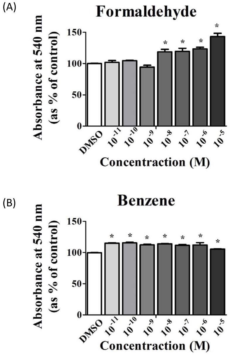
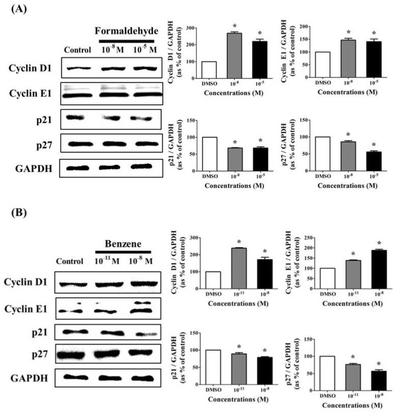
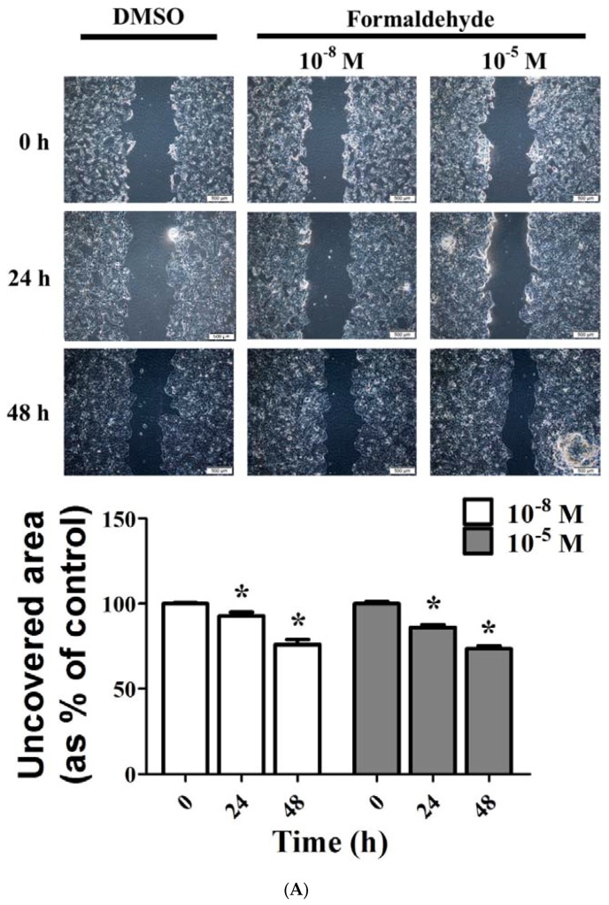
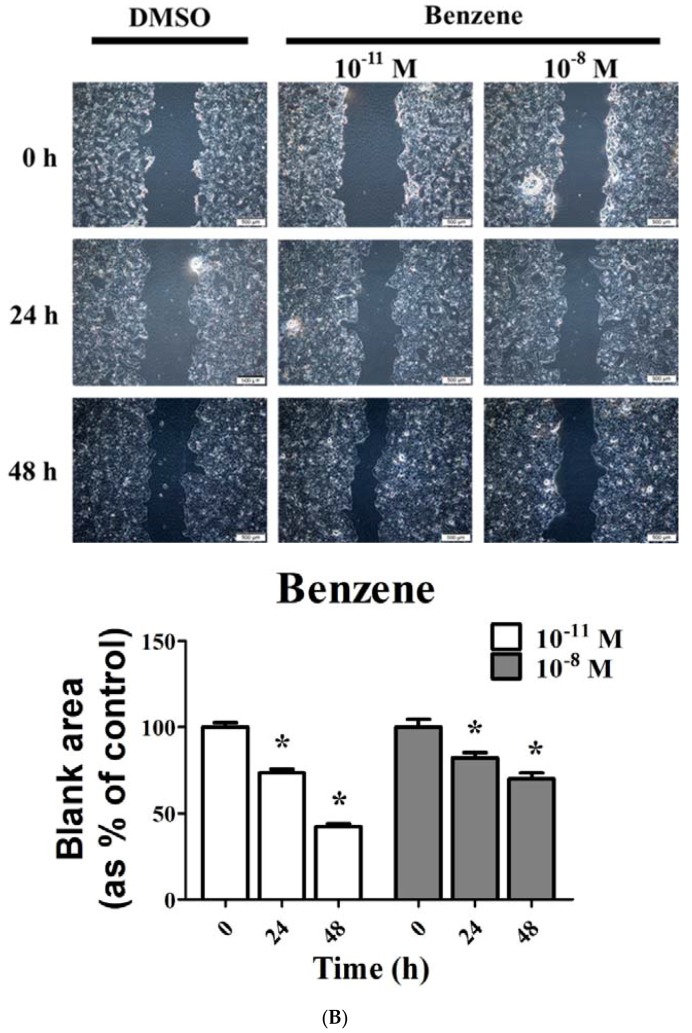
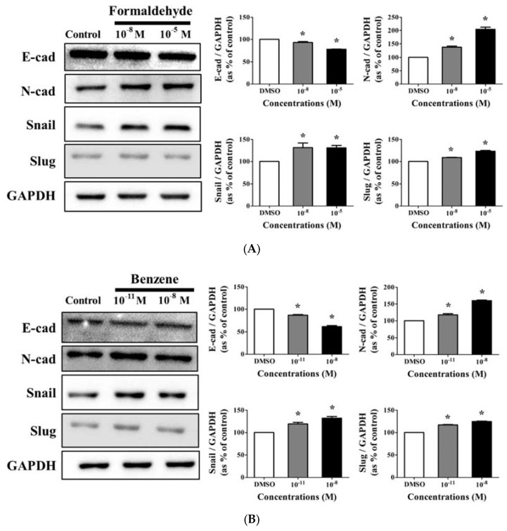
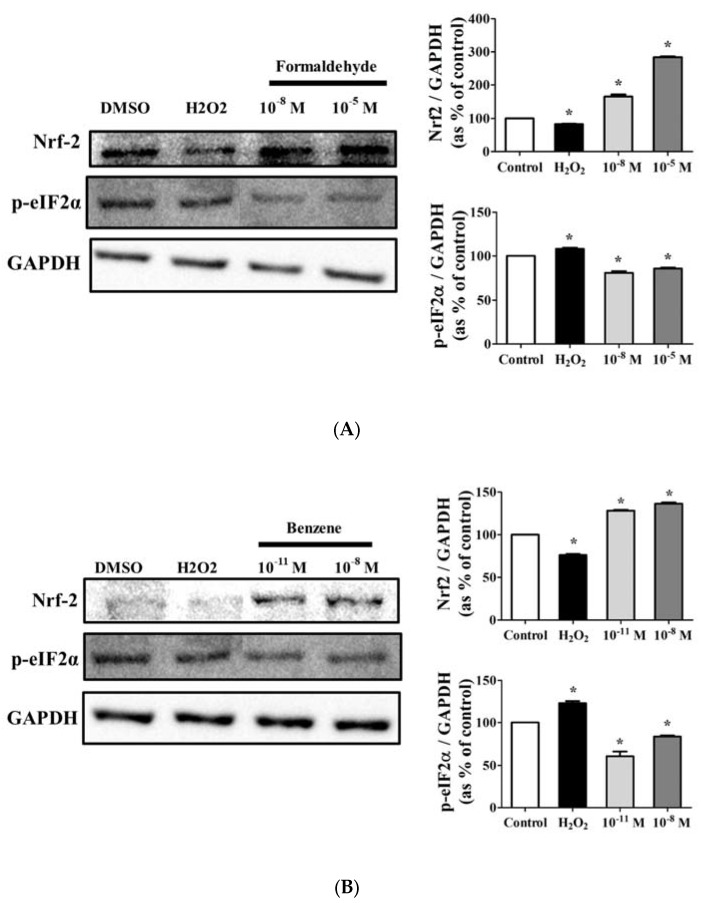
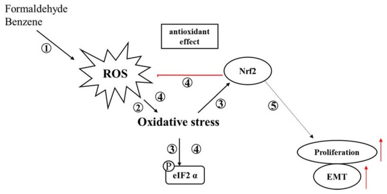
Cigarette smoke (CS) causes about 480,000 deaths each year worldwide, and it is well-known to have harmful effects on the human body, leading to heart disease, stroke, lung cancer, and cardiovascular problems. In this study, the effects of formaldehyde (FA) and benzene (Bz), the main components of CS, on cell proliferation and epithelial mesenchymal transition (EMT) of JEG-3 human choriocarcinoma cells were examined to confirm the relationship between CS components and placenta carcinoma. Upon MTT assay, FA (10-8 M to 10-5 M) and Bz (10-11 M to 10-8 M) increased JEG-3 cell proliferation. Western blot assay revealed that the protein expression of cyclin D1 and E1 increased, while the levels of p21 and p27 were reduced following treatment. In Scratch assay, FA (10-8 M and 10-5 M) and Bz (10-11 M and 10-8 M) increased migration of JEG-3 cells at 24 h and 48 h compared with that at 0 h. In addition, the expression of the epithelial marker, E-cadherin, was significantly decreased, while the expression of the mesenchymal marker, N-cadherin, was significantly increased by FA (10-8 M and 10-5 M) and Bz (10-11 M and 10-8 M). snail and slug transcriptional factors were associated with EMT, which were also up-regulated by FA and Bz, indicating that FA and Bz lead to an increase in the EMT process in JEG-3 choriocarcinoma cells. We further evaluated reactive oxygen species (ROS) and activation of antioxidant effect using dichlorofluorescin diacetate (DCFH-DA) and Western blot assay. FA and Bz increased the ROS production and an antioxidant related marker, Nrf2, in JEG-3 cells. However, eIF2α levels were reduced by FA and Bz via activation of the antioxidant reaction. Taken together, these results indicated that FA and Bz induce the growth and migration of human choriocarcinoma cells via regulation of the cell cycle and EMT and activation of ROS and antioxidant related markers.

Keywords: EMT; benzene; cell cycle; cigarette smoke; formaldehyde; placenta choriocarcinoma.


Conflict of interest statement

The authors declare no conflict of interest.

Figures

Figure 1
Figure 1
Effect of cigarette smoke components on the proliferation of human placenta carcinoma cells JEG-3. JEG-3 cells were seeded at 5000 cell/well in 96-well plates. After 1 day of incubation, medium treated with (A) formaldehyde (FA) (10−8 M to 10−5 M), (B) benzene (Bz) (10−11 M to 10−8 M), or 0.1 % dimethyl sulfoxide (DMSO) (a control) was added for 9 days. The cell proliferation was then evaluated through MTT assay. Values shown are the means ± SD. * mean values were significantly different from control, p < 0.05. (Dunnett’s multiple comparison test).
Figure 2
Figure 2
Effect of FA and Bz on protein expression of cell cycle related genes in JEG-3 cells. JEG-3 cells were seeded in 100 mm dishes and treated with medium containing DMSO (control), (A) FA (10−11 M to 10−5 M), or (B) Bz (10−11 M to 10−5 M) for 72 h. After protein extraction, Western blot assay was conducted to conform to the protein expression of cell cycle related genes (cyclin D1, cyclin E1), cell cycle arrest genes (p21 and p27), and housekeeping genes (glyceraldehyde 3-phosphate dehydrogenase (GAPDH)). Quantification of cyclin D1, cyclin E1, p21, and p27 protein was conducted by measuring band densities using a CS analyzer 4 (ATTO, Corp., Japan), and their protein levels were normalized by the band value of GAPDH. Values shown are the means ± SD. * mean values were significantly different from 0.1% DMSO (control), p < 0.05. (Dunnett’s multiple comparison test).
Figure 3
Figure 3
Effect of FA and Bz on migration activity of JEG-3 cells. JEG-3 cells were seeded in 6-well plates at 80% density, after which a region was scratched with the same length and width. Samples were then treated with medium containing 0.1% DMSO (control), (A) FA (10−8 M and 10−5 M), or (B) Bz (10−11 M and 10−8 M) for 48 h. Images shown were taken under a microscope at 40× magnification. Quantification of migration activity was conducted using a Cellsens dimension program. Values shown are the means ± SD. * mean values were significantly different from control, p < 0.05. (Dunnett’s multiple comparison test).
Figure 3
Figure 3
Effect of FA and Bz on migration activity of JEG-3 cells. JEG-3 cells were seeded in 6-well plates at 80% density, after which a region was scratched with the same length and width. Samples were then treated with medium containing 0.1% DMSO (control), (A) FA (10−8 M and 10−5 M), or (B) Bz (10−11 M and 10−8 M) for 48 h. Images shown were taken under a microscope at 40× magnification. Quantification of migration activity was conducted using a Cellsens dimension program. Values shown are the means ± SD. * mean values were significantly different from control, p < 0.05. (Dunnett’s multiple comparison test).
Figure 4
Figure 4
Effect of FA and Bz on protein expression of epithelial mesenchymal transition (EMT) markers in JEG-3. JEG-3 cells were seeded in 100 mm dishes and treated medium with 0.1% DMSO (control), (A) FA (10−8 M and 10−5 M), or (B) Bz (10−11 M to and 10−8 M) for 72 h. After protein extraction, Western blot was conducted to confirm the protein expression of epithelial marker (E-cadherin), mesenchymal markers (N-cadherin, Snail and Slug), and the housekeeping gene (GAPDH). Quantification of E-cadherin, N-cadherin, Snails and Slug protein was conducted by measuring the band densities using a CS analyzer 4 (ATTO, Corp., Japan), and their protein levels were normalized by the band value of GAPDH. Values shown are the means ± SD. * mean values were significantly different from control, p < 0.05. (Dunnett’s multiple comparison test).
Figure 5
Figure 5
Activation of reactive oxygen species (ROS) by treatment with FA and Bz. JEG-3 cells were seeded at 2 × 10⁴ cell per well in a 96-well plate. After 1 day of incubation, medium treated with (A) FA (10−8 M and 10−5 M), (B) Bz (10−11 M and 10−8 M), or 0.1% DMSO (a control) was added for 72 h, and 3% H2O2 was treated for 30 min as a positive control to induce ROS. The culture medium was then removed, and each well was treated with 200 µL 20,70-dichlorofluorescein diacetate (DCF-DA) solution for 30 min. To detect ROS activation, fluorescence intensity was measured using an IX70 fluorescence microscope (Olympus, Japan) in a dark room at room temperature.
Figure 6
Figure 6
Effect of FA and Bz on protein expression of antioxidant factor nuclear factor erythroid 2 (NFE2)-related factor 2 (Nrf2) and endoplasmic reticulum (ER) stress marker eIF2α in JEG-3. JEG-3 cells were seeded in 100 mm dishes and treated with medium containing 0.1% DMSO (control), (A) FA (10−11 M and 10−8 M), or (B) Bz (10−8 M and 10−5 M) for 72 h. After protein extraction, Western blot was conducted to confirm the protein expression of the antioxidant factor, Nrf2, the ER stress marker, eIF2α, and the housekeeping gene, GAPDH. Quantification of Nrf2 and eIF2α protein was conducted by measuring band densities using a CS analyzer 4 (ATTO, Corp., Japan), and their protein levels were then normalized by the band value of GAPDH. Values shown are the means ± SD. * mean values were significantly different from 0.1% DMSO (control), p < 0.05. (Dunnett’s multiple comparison test).
Figure 7
Figure 7
Roles of FA and Bz in the exhibition of cell proliferation and the promotion of EMT or in the prevention of apoptosis. ① FA and Bz activate the intracellular ROS of JEG-3, ② which in turn activates oxidative stress. ③ This activates apoptosis by activating apoptosis-related eIF2, or by activating the antioxidant factor, Nrf2, ④ to regulate the activation of ROS by inducing an antioxidant effect through the increase of Nrf2 which blocks the activation of ROS. However, FA and Bz not only activate ROS, but also increase the activity of Nrf2, which prevents the activation of oxidative stress and apoptosis. ⑤ In addition, FA and Bz induce cell proliferation and EMT, either directly or indirectly through Nrf2. Therefore, FA and Bz inhibit apoptosis through the ROS-Nrf2 pathway, leading to an increase in proliferation and EMT.

Similar articles

Cited by

References

    1. Sobus S.L., Warren G.W. The biologic effects of cigarette smoke on cancer cells. Cancer. 2014;120:3617–3626. doi: 10.1002/cncr.28904. - DOI - PubMed
    1. Newcomb P.A., Carbone P.P. The health consequences of smoking: Cancer. Med. Clin. N. Am. 1992;76:305–331. doi: 10.1016/S0025-7125(16)30355-8. - DOI - PubMed
    1. Hanaki T., Horikoshi Y., Nakaso K., Nakasone M., Kitagawa Y., Amisaki M., Arai Y., Tokuyasu N., Sakamoto T., Honjo S., et al. Nicotine enhances the malignant potential of human pancreatic cancer cells via activation of atypical protein kinase C. Biochim. Biophys. Acta. 2016;1860(Pt A11):2404–2415. doi: 10.1016/j.bbagen.2016.07.008. - DOI - PubMed
    1. Kim C.W., Go R.E., Lee H.M., Hwang K.A., Lee K., Kim B., Lee M.Y., Choi K.C. Cigarette smoke extracts induced the colon cancer migration via regulating epithelial mesenchymal transition and metastatic genes in human colon cancer cells. Environ. Toxicol. 2016;32:690–704. doi: 10.1002/tox.22271. - DOI - PubMed
    1. Pesch B., Kendzia B., Gustavsson P., Jockel K.H., Johnen G., Pohlabeln H., Olsson A., Ahrens W., Gross I.M., Bruske I., et al. Cigarette smoking and lung cancer-relative risk estimates for the major histological types from a pooled analysis of case-control studies. Int. J. Cancer. 2012;131:1210–1219. doi: 10.1002/ijc.27339. - DOI - PMC - PubMed

LinkOut - more resources