Review
. 2017 Jul 30;8(3):32.
doi: 10.3390/jfb8030032.

Potential New Non-Invasive Therapy Using Artificial Oxygen Carriers for Pre-Eclampsia

Affiliations
Review

Potential New Non-Invasive Therapy Using Artificial Oxygen Carriers for Pre-Eclampsia

Hidenobu Ohta et al. J Funct Biomater. .

Abstract

The molecular mechanisms of pre-eclampsia are being increasingly clarified in animals and humans. With the uncovering of these mechanisms, preventive therapy strategies using chronic infusion of adrenomedullin, vascular endothelial growth factor-121 (VEGF-121), losartan, and sildenafil have been proposed to block narrow spiral artery formation in the placenta by suppressing related possible factors for pre-eclampsia. However, although such preventive treatments have been partly successful, they have failed in ameliorating fetal growth restriction and carry the risk of possible side-effects of drugs on pregnant mothers. In this study, we attempted to develop a new symptomatic treatment for pre-eclampsia by directly rescuing placental ischemia with artificial oxygen carriers (hemoglobin vesicles: HbV) since previous data indicate that placental ischemia/hypoxia may alone be sufficient to lead to pre-eclampsia through up-regulation of sFlt-1, one of the main candidate molecules for the cause of pre-eclampsia. Using a rat model, the present study demonstrated that a simple treatment using hemoglobin vesicles for placental ischemia rescues placental and fetal hypoxia, leading to appropriate fetal growth. The present study is the first to demonstrate hemoglobin vesicles successfully decreasing maternal plasma levels of sFlt-1 and ameliorating fetal growth restriction in the pre-eclampsia rat model (p < 0.05, one-way ANOVA). In future, chronic infusion of hemoglobin vesicles could be a potential effective and noninvasive therapy for delaying or even alleviating the need for Caesarean sections in pre-eclampsia.

Keywords: Keywords: pre-eclampsia; brain damage; fetus; hemoglobin vesicle; hypoxic condition; placenta.


Conflict of interest statement

The authors declare no conflict of interest

Figures

Figure 1
Figure 1
The safety and placental transfer of hemoglobin vesicles (HbV) to fetus in pregnancy. (a) Time course of the gains in body weights of pregnant mother rats before and after daily repeated infusions (DRI) of HbV or saline for 7 days at a dose rate of 2 mL/kg/day (n = 5 for each group; value: average ± s.d.). The weights of fetuses (b) and the placentas (c) after 7 days’ DRI of HbV or saline (n = 5 for each group; value: average ± s.d.). (d) Tissue distributions of 125I-HbV at 12 h after administration to pregnant rats. Rats received a single injection of 125I-HbV from the tail vein at a dose of 1400 mg/kg. Twelve hours after injection, each organ was collected. (n = 5 for each group; value: average ± s.d.).
Figure 2
Figure 2
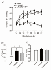
Effects of hemoglobin vesicle (HbV) infusion on maternal blood pressure and sFlt-1/sEng production in pregnant rats. (a) Chronological changes in systolic blood pressure during pregnancy in saline (control), NG-nitro-l-arginine methyl ester (l-NAME)-only treated and l-NAME + HbV treated pregnant rats. Closed circles, saline control pregnant rats; open circles, l-NAME-only treated pregnant rats; closed triangles, l-NAME + HbV treated pregnant rats. The data for each group are expressed as mean values ± s.e. (n = 5). Significant difference between control and l-NAME-only treated rats or l-NAME + HbV treated rats was observed (* p < 0.05, two-way ANOVA, Dunette); (b)The plasma sFlt-1 levels in l-NAME-only treated rats were significantly higher compared with those in saline control pregnant rats or l-NAME + HbV treated rats; (c)There was no statistical significance in the plasma sEng among the three groups. Data are expressed as mean ± s.e. (* p < 0.05, one-way ANOVA, Dunette).
Figure 3
Figure 3
Effects of hemoglobin vesicle (HbV) infusion on placental hypoxia in pregnant rats. Hypoxic-inducible factor 1α (HIF-1α) (brown signal) shows stronger signal activity in the labyrinth and spongiotrophoblast of the l-NAME-only treated group (b,e) compared with that of the saline control group (a,d) or the l-NAME + HbV treated group (c,f). Arrows in (b) indicate representative HIF-1α positive cells (dark brown cells). Scale bar: 300 µm. Quantification of the HIF-1α-positive cells in the labyrinth (g) and spongiotrophoblast (h). Western blot analysis of HIF-1α in the placental tissues from saline, l-NAME, and l-NAME + HbV treated pregnant rats (i).
Figure 4
Figure 4
Effects of hemoglobin vesicle (HbV) infusion on fetal hypoxia. Photon flux (p/s/cm2/sr) from the heterozygous Rosa 26::luc fetuses is displayed according to the scale bar at the right side. Compared to the basal state of saline injection (a), 60 s exposures show that light emission increased after an acute HbV injection (b). Quantification of the HIF-1α-positive cells in the cortex (c) and hippocampus (d). Data are expressed as mean ± s.e. (* p < 0.05, one-way ANOVA, Dunette). Maternal l-NAME injection induced fetal brain hypoxia, as indicated by more extensive HIF-1α positive staining at the cortex and hippocampus in fetal brain compared to the same areas of fetal brains from saline or l-NAME + HbV treated mothers. Note that HbV attenuated l-NAME-induced fetal brain hypoxia.
Figure 5
Figure 5
Effects of hemoglobin vesicle (HbV) infusion on fetal brain and body growth under hypoxic conditions. The hippocampus of G21 fetuses from the pregnant rats treated with saline (a, n = 5), l-NAME (b, n = 5), or l-NAME + HbV (c, n = 5) are shown. The glial fibrillary acidic protein (GFAP) positive staining in the hippocampus of fetuses (b) from l-NAME treated mothers was stronger compared with those from the saline treated mothers (a) or l-NAME + HbV treated mothers (c). This indicated that maternal l-NAME injection induced fetal brain astrogliosis and HbV reduced the l-NAME-induced fetal brain damage. Quantification of the GFAP-positive area in the hippocampus (g). Data are expressed as mean ± s.e. (* p < 0.05, one-way ANOVA, Dunette). The number of NeuN-positive cells in the hippocampus of fetuses (e) from l-NAME treated mothers was smaller compared with that in the saline treated mothers (d) or l-NAME + HbV treated mothers (f). This indicated that maternal l-NAME injection induced neural damage in the fetal brain while HbV reduced the l-NAME-induced fetal brain damage. Quantification of the NeuN-positive cells in the hippocampus (h). Data are expressed as mean ± s.e. (* p < 0.05, one-way ANOVA, Dunette). The weight gains of rat fetuses (i) and the placentas (j) after chronic HbV or saline infusion at a dose rate of 2 mL/kg/day (n = 5 for each group; value: average ± s.e.) for 7 days. Note that HbV rescued the fetal and placental weights in l-NAME + HbV treated groups compared those in l-NAME-only treated groups. Scale bar: 100 µm.

Similar articles

Cited by

References

    1. Sibai B., Dekker G., Kupferminc M. Pre-eclampsia. Lancet. 2005;365:785–799. - PubMed
    1. Bibbins-Domingo K., Grossman D.C., Curry S.J., Barry M.J., Davidson K.W., Doubeni C.A., Epling J.W., Kemper A.R., Krist A.H., Kurth A.E., et al. Screening for preeclampsia: US Preventive Services Task Force recommendation statement. JAMA. 2017;317:1661–1667. - PubMed
    1. Henderson J.T., Thompson J.H., Burda B.U., Cantor A. Preeclampsia screening: Evidence report and systematic review for the US Preventive Services Task Force. JAMA. 2017;317:1668–1683. doi: 10.1001/jama.2016.18315. - DOI - PubMed
    1. Myatt L., Webster R.P. Vascular biology of preeclampsia. J. Thromb. Haemost. 2009;7:375–384. - PubMed
    1. Powe C.E., Levine R.J., Karumanchi S.A. Preeclampsia, a disease of the maternal endothelium: The role of antiangiogenic factors and implications for later cardiovascular disease. Circulation. 2011;123:2856–2869. doi: 10.1161/CIRCULATIONAHA.109.853127. - DOI - PMC - PubMed

LinkOut - more resources