. 2017 Jul 31;18(8):1667.
doi: 10.3390/ijms18081667.

Tricetin Induces Apoptosis of Human Leukemic HL-60 Cells through a Reactive Oxygen Species-Mediated c-Jun N-Terminal Kinase Activation Pathway

Affiliations

Tricetin Induces Apoptosis of Human Leukemic HL-60 Cells through a Reactive Oxygen Species-Mediated c-Jun N-Terminal Kinase Activation Pathway

Ming-Hsien Chien et al. Int J Mol Sci. .

Abstract

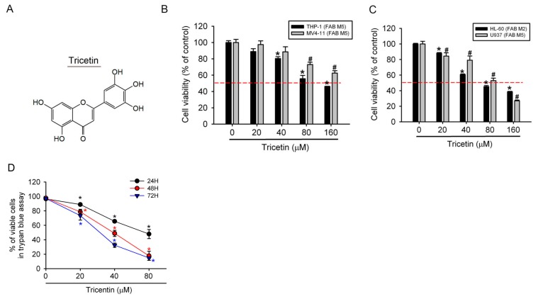
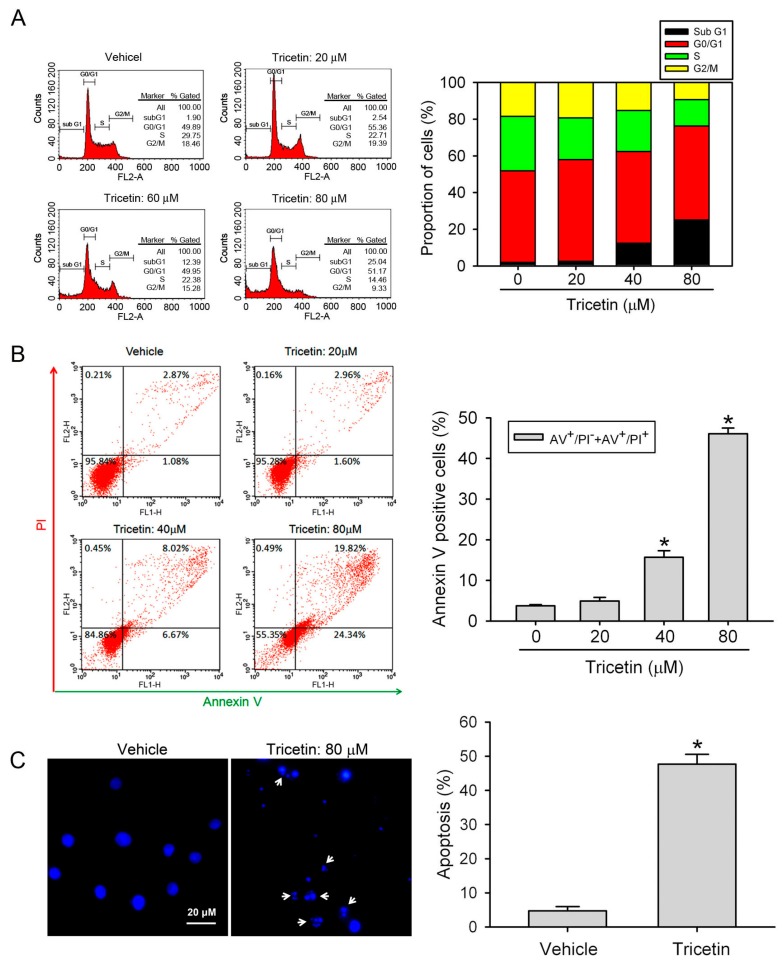
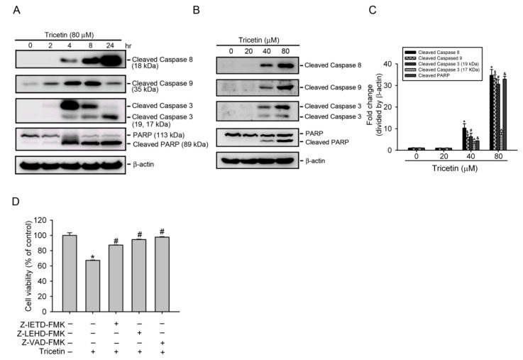
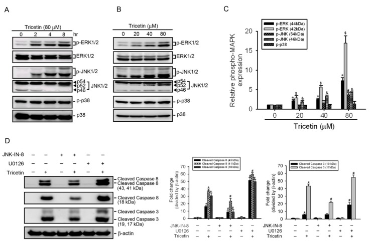
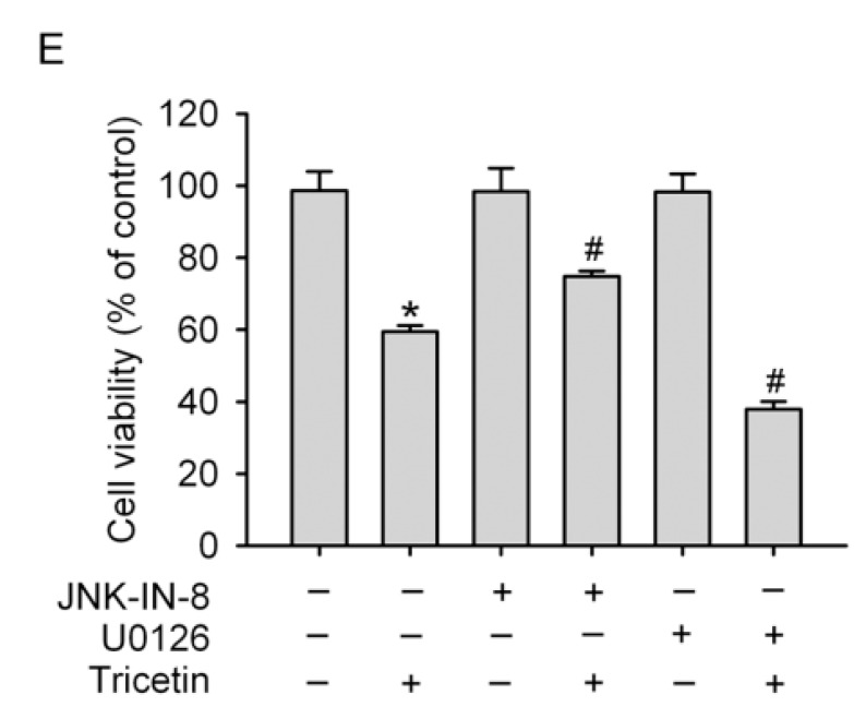
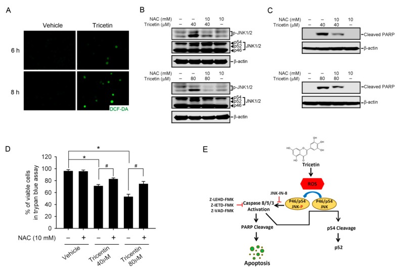
Tricetin is a dietary flavonoid with cytostatic properties and antimetastatic activities in various solid tumors. The anticancer effect of tricetin in nonsolid tumors remains unclear. Herein, the molecular mechanisms by which tricetin exerts its anticancer effects on acute myeloid leukemia (AML) cells were investigated. Results showed that tricetin inhibited cell viability in various types of AML cell lines. Tricetin induced morphological features of apoptosis such as chromatin condensation and phosphatidylserine (PS) externalization, and significantly activated proapoptotic signaling including caspase-8, -9, and -3 activation and poly(ADP-ribose) polymerase (PARP) cleavage in HL-60 AML cells. Of note, tricetin-induced cell growth inhibition was dramatically reversed by a pan caspase and caspase-8- and -9-specific inhibitors, suggesting that this compound mainly acts through a caspase-dependent pathway. Moreover, treatment of HL-60 cells with tricetin induced sustained activation of extracellular signal-regulated kinase (ERK) and c-Jun N-terminal kinase (JNK), and inhibition of ERK and JNK by their specific inhibitors respectively promoted and abolished tricetin-induced cell apoptosis. Dichlorofluorescein (DCF) staining showed that intracellular reactive oxygen species (ROS) levels were higher in tricetin-treated HL-60 cells compared to the control group. Moreover, an ROS scavenger, N-acetylcysteine (NAC), reversed tricetin-induced JNK activation and subsequent cell apoptosis. In conclusion, our results indicated that tricetin induced cell death of leukemic HL-60 cells through induction of intracellular oxidative stress following activation of a JNK-mediated apoptosis pathway. A combination of tricetin and an ERK inhibitor may be a better strategy to enhance the anticancer activities of tricetin in AML.

Keywords: acute myeloid leukemia; apoptosis; c-Jun N-terminal kinase; reactive oxygen species; tricetin.


Conflict of interest statement

The authors declare no conflict of interest.

Figures

Figure 1
Figure 1
Tricetin treatment results in reduced cell viability of human acute myeloid leukemia (AML) cell lines. (A) The chemical structure of tricetin; (B,C) Four human AML cell lines (HL-60, U937, THP-1, and MV4-11) were treated with the vehicle (DMSO) or tricetin (0~160 μM) in serum-containing medium for 24 h; (D) HL-60 cells were treated with different concentrations of tricetin (0~80 μM) for 24, 48, and 72 h. Cell viability was determined by a trypan blue exclusion assay. Results are expressed as multiples of cell viability. Values represent the mean ± standard error (SE) of three independent experiments. *, # p < 0.05, compared to the vehicle groups. The dashed line indicates 50% growth inhibition of cell viability.
Figure 2
Figure 2
Effect of tricetin on HL-60 cell-cycle regulation and apoptosis. (A) HL-60 cells were treated with different concentrations of tricetin (0~80 μM) for 24 h. The cell-cycle phase distribution and cell death in the sub-G1 phase were analyzed by fluorescence-activated cell sorting (FACS) after propidium iodide (PI) staining. Data are shown as the cell-cycle distribution profile by FACS and the percentage distribution of cells in the sub-G1, G0/G1, S, and G2/M phases; (B) HL-60 cells were treated with different concentrations of tricetin (0~80 μM) for 24 h. Quantitative analysis of cell apoptosis by FACS after staining with Annexin-V and PI. In the dot plots, percentages of Annexin-V+/PI (cells in early apoptosis, bottom right quadrant) and Annexin-V+/PI+ (cells in late apoptosis, top right quadrant) are shown (C) HL-60 cells were treated with 80 μM tricetin for 24 h and analyzed by fluorescence microscopy after 4′,6-diamidino-2-phenylindole (DAPI) staining. White arrows indicate apoptotic HL-60 cells. Percentage of apoptotic cells expresses ratio of apoptotic cells to the total cell number. For each sample 200 cells were assessed. Data are expressed as the mean ± SE of three independent experiments. * p < 0.05, compared to the vehicle group.
Figure 3
Figure 3
Tricetin induces caspase-dependent apoptotic cell death in HL-60 cells. (A) Expression levels of cleaved caspases-3, -8, and -9, and poly(ADP-ribose) polymerase (PARP) were assessed by a Western blot analysis after treatment with 80 μM tricetin for indicated time points; (B) Activated caspase-8, -9, and -3, and cleaved PARP protein expressions were upregulated in a concentration-dependent fashion after treatment of HL-60 cells with various concentrations of tricetin (0~80 μM) for 8 h; (C) Quantitative results of cleaved caspase-3, -8, and -9, and PARP protein levels, which were adjusted to the β-actin protein level and expressed as multiples of induction beyond each respective control. Values are presented as the mean ± SE of three independent experiments. *, #, &, ^, $ p < 0.05, compared to the vehicle control groups; (D) Cells were treated with 40 μM tricetin for 24 h in the presence or absence of 50 μM Z-VAD-FMK, Z-LEHD-FMK, or Z-IETD-FMK. Cell proliferation was determined by an MTS assay. Data are presented as the mean ± SE of three independent experiments performed in triplicate. * p < 0.05, control vs. tricetin; # p < 0.05, tricetin vs. Z-VAD-FMK, Z-LEHD-FMK, or Z-IETD-FMK plus tricetin.
Figure 4
Figure 4
Role of mitogen-activated protein kinases (MAPKs) in tricetin-induced apoptotic cell death in HL-60 cells. (A,B) Phosphorylation levels of extracellular signal-regulated kinase (ERK)1/2, p38, and c-Jun N-terminal kinase (JNK)1/2 were assessed by a Western blot analysis after treatment of HL-60 cells with 80 μM tricetin for indicated time points (A) or with various concentrations of tricetin (0~80 μM) for 8 h (B,C) Quantitative results of phopho-ERK1/2, p38, and JNK1/2 protein levels, which were adjusted to the total ERK1/2, p38, and JNK1/2 protein levels and expressed as multiples of induction beyond each respective control. Values are presented as the mean ± SE of three independent experiments. *, #, &, $ p < 0.05, compared to the vehicle control groups; (D,E) HL-60 cells were pretreated with or without 5 μM U0126 or 1 μM JNK-IN-8 for 1 h followed by tricetin (40 μM) treatment for an additional 24 h. Expression levels of cleaved caspases-3, -8, and cell viability were respectively determined by a Western blot analysis (D, left panel) and a CCK-8 assay (E). Quantitative results of cleaved caspase-3 and -8 protein levels, which were adjusted to the β-actin protein level and expressed as multiples of induction beyond each respective control (D, right panel). Values represent the mean ± SE of three independent experiments. * p < 0.05, control vs. tricetin; # p < 0.05, tricetin vs. U0126 or JNK-IN-8 plus tricetin.
Figure 4
Figure 4
Role of mitogen-activated protein kinases (MAPKs) in tricetin-induced apoptotic cell death in HL-60 cells. (A,B) Phosphorylation levels of extracellular signal-regulated kinase (ERK)1/2, p38, and c-Jun N-terminal kinase (JNK)1/2 were assessed by a Western blot analysis after treatment of HL-60 cells with 80 μM tricetin for indicated time points (A) or with various concentrations of tricetin (0~80 μM) for 8 h (B,C) Quantitative results of phopho-ERK1/2, p38, and JNK1/2 protein levels, which were adjusted to the total ERK1/2, p38, and JNK1/2 protein levels and expressed as multiples of induction beyond each respective control. Values are presented as the mean ± SE of three independent experiments. *, #, &, $ p < 0.05, compared to the vehicle control groups; (D,E) HL-60 cells were pretreated with or without 5 μM U0126 or 1 μM JNK-IN-8 for 1 h followed by tricetin (40 μM) treatment for an additional 24 h. Expression levels of cleaved caspases-3, -8, and cell viability were respectively determined by a Western blot analysis (D, left panel) and a CCK-8 assay (E). Quantitative results of cleaved caspase-3 and -8 protein levels, which were adjusted to the β-actin protein level and expressed as multiples of induction beyond each respective control (D, right panel). Values represent the mean ± SE of three independent experiments. * p < 0.05, control vs. tricetin; # p < 0.05, tricetin vs. U0126 or JNK-IN-8 plus tricetin.
Figure 5
Figure 5
Tricetin-induced intracellular oxidative stress as an initial signal for c-Jun N-terminal kinase (JNK)-mediated apoptosis in HL-60 cells. (A) HL-60 cells were treated with 80 μM tricetin for the indicated time, and then the total reactive oxygen species (ROS) level was measured by H2DCF-DA staining under a fluorescence microscope. Original magnification, 200×; (B,C) HL-60 cells were pretreated with or without 10 mM N-acetylcysteine (NAC) for 1 h followed by treatment with 40 or 80 μM tricetin for 8 h; levels of JNK1/2, p-JNK1/2, cleaved poly(ADP ribose) polymerase (PARP), and β-actin were detected by a Western blot analysis; (D) HL-60 cells were pretreated with or without 10 mM NAC for 1 h followed by treatment with 40 or 80 μM tricetin for 12 h Trypan blue exclusion assay was used to quantify the cell viability change in each group. Values represent the mean ± SE of three independent experiments. * p < 0.05, control vs. tricetin; # p < 0.05, tricetin vs. NAC plus tricetin; (E) Proposed signal transduction pathways by which tricetin induces apoptosis of acute myeloid leukemias (AML) cells. The antileukemic activity of tricetin was attributed to its apoptosis induction by increasing ROS production and further inducing activation of JNK and caspases-8, -9, and -3. The p54 JNK cleavage was also induced by tricetin-mediated ROS upregulation. Black arrows indicate induced effects and red t-bar indicate inhibitory effects.

Similar articles

Cited by

References

    1. Burnett A.K., Hills R.K., Milligan D.W., Goldstone A.H., Prentice A.G., McMullin M.F., Duncombe A., Gibson B., Wheatley K. Attempts to optimize induction and consolidation treatment in acute myeloid leukemia: Results of the MRC AML12 trial. J. Clin. Oncol. 2010;28:586–595. doi: 10.1200/JCO.2009.22.9088. - DOI - PubMed
    1. Tallman M.S., Gilliland D.G., Rowe J.M. Drug therapy for acute myeloid leukemia. Blood. 2005;106:1154–1163. doi: 10.1182/blood-2005-01-0178. - DOI - PubMed
    1. Estey E., Dohner H. Acute myeloid leukaemia. Lancet. 2006;368:1894–1907. doi: 10.1016/S0140-6736(06)69780-8. - DOI - PubMed
    1. Shanafelt T.D., Lee Y.K., Call T.G., Nowakowski G.S., Dingli D., Zent C.S., Kay N.E. Clinical effects of oral green tea extracts in four patients with low grade B-cell malignancies. Leuk. Res. 2006;30:707–712. doi: 10.1016/j.leukres.2005.10.020. - DOI - PubMed
    1. Lucas D.M., Still P.C., Perez L.B., Grever M.R., Kinghorn A.D. Potential of plant-derived natural products in the treatment of leukemia and lymphoma. Curr. Drug Targets. 2010;11:812–822. doi: 10.2174/138945010791320809. - DOI - PMC - PubMed

MeSH terms

LinkOut - more resources