. 2017 Sep 18;30(9):1720-1729.
doi: 10.1021/acs.chemrestox.7b00172. Epub 2017 Aug 20.

Variation in Extracellular Detoxification Is a Link to Different Carcinogenicity among Chromates in Rodent and Human Lungs

Affiliations

Variation in Extracellular Detoxification Is a Link to Different Carcinogenicity among Chromates in Rodent and Human Lungs

Casey Krawic et al. Chem Res Toxicol. .

Abstract

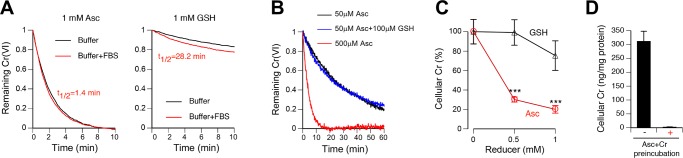
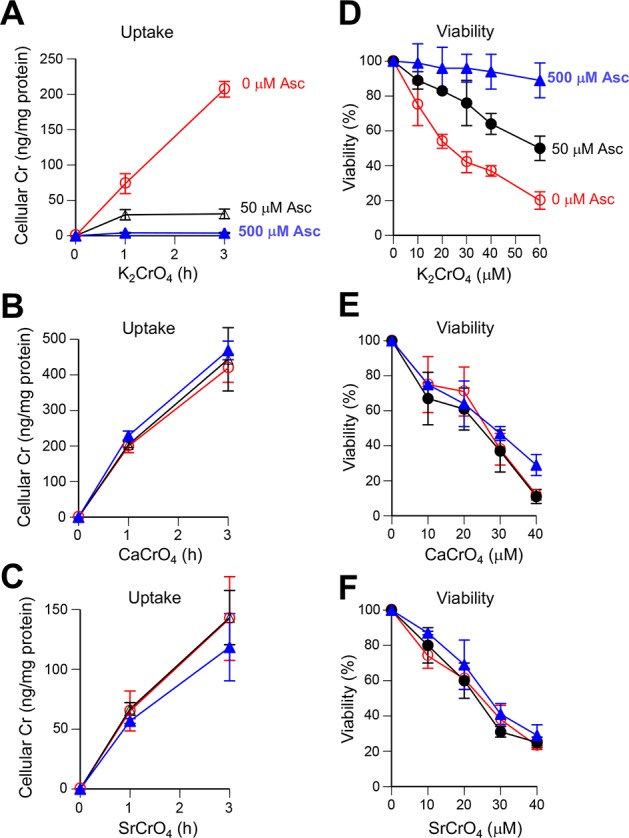
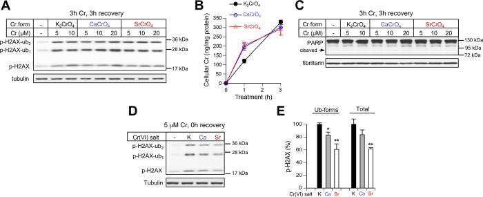
Inhalation of soluble chromium(VI) is firmly linked with higher risks of lung cancer in humans. However, comparative studies in rats have found a high lung tumorigenicity for moderately soluble chromates but no tumors for highly soluble chromates. These major species differences remain unexplained. We investigated the impact of extracellular reducers on responses of human and rat lung epithelial cells to different Cr(VI) forms. Extracellular reduction of Cr(VI) is a detoxification process, and rat and human lung lining fluids contain different concentrations of ascorbate and glutathione. We found that reduction of chromate anions in simulated lung fluids was principally driven by ascorbate with only minimal contribution from glutathione. The addition of 500 μM ascorbate (∼rat lung fluid concentration) to culture media strongly inhibited cellular uptake of chromate anions and completely prevented their cytotoxicity even at otherwise lethal doses. While proportionally less effective, 50 μM extracellular ascorbate (∼human lung fluid concentration) also decreased uptake of chromate anions and their cytotoxicity. In comparison to chromate anions, uptake and cytotoxicity of respirable particles of moderately soluble CaCrO4 and SrCrO4 were much less sensitive to suppression by extracellular ascorbate, especially during early exposure times and in primary bronchial cells. In the absence of extracellular ascorbate, chromate anions and CaCrO4/SrCrO4 particles produced overall similar levels of DNA double-stranded breaks, with less soluble particles exhibiting a slower rate of breakage. Our results indicate that a gradual extracellular dissolution and a rapid internalization of calcium chromate and strontium chromate particles makes them resistant to detoxification outside the cells, which is extremely effective for chromate anions in the rat lung fluid. The detoxification potential of the human lung fluid is significant but much lower and insufficient to provide a threshold-type dose dependence for soluble chromates.


Conflict of interest statement

The authors declare no competing financial interest.

Figures

Figure 1
Figure 1
Impact of ascorbate (Asc) and glutathione (GSH) on reduction and cellular uptake of chromate anion. All reactions used solubilized K2CrO4 as a source of chromate anions. (A) Time-course of 50 μM chromate reduction at 37 °C (buffer, 50 mM HEPES, 100 mM NaCl, pH 7.0; buffer+FBS, HEPES buffer containing 10% fetal bovine serum). Data are means of triplicate measurements. Error bars were smaller than 5% of the mean and are not shown for clarity. (B) Chromate reduction in HEPES buffer by reducer concentrations that are typically found in lung lining fluids. Reactions contained 5 μM Cr(VI). Data are means of triplicate measurements. (C) Uptake of chromate by H460 cells in the presence of extracellular Asc or GSH. Cells were incubated with 10 μM Cr(VI) for 1 h. Data are means ± SD, n = 3, ∗∗∗p < 0.001 relative to 0 mM reducer. (D) Elimination of Cr accumulation by a preincubation of 10 μM chromate in culture media with 1 mM Asc (1 h, 37 °C) prior to the addition to H460 cells. Cells were incubated with Cr-containing media for 1 h. Data are means ± SD, n = 3.
Figure 2
Figure 2
Uptake of chromate anions and CaCrO4 particles by human H460 cells. Cells were incubated for 1 h with 5 μM Cr(VI) in the form of solubilized K2CrO4 or CaCrO4 particles (0.8% final ethanol concentration). (A) Cellular Cr levels after incubations with soluble and particulate Cr(VI) in the presence of 0, 50, and 500 μM Asc in media. Data are means ± SD, n = 3, ∗p < 0.05, ∗∗∗p < 0.001 relative to 0 μM Asc. (B) Cellular Cr accumulation from soluble and particulate Cr(VI) in the presence of extracellular 50 μM Asc with and without 100 μM GSH. Data are means ± SD, n = 3. (C) Cellular amounts of Cr at 0 and 3 h postexposure. Data are means ± SD, n = 3.
Figure 3
Figure 3
Effects of ethanol on Cr(VI) metabolism and cell viability. (A) Kinetics of chromate reduction by Asc in the presence of ethanol. Reactions contained 0–2% ethanol, 200 μM Asc, 20 μM chromate in HEPES-NaCl buffer, pH 7.0. Data are means of triplicate measurements. (B) Uptake of chromate anions (solubilized K2CrO4) and CaCrO4 particles by H460 cells. Cells were incubated with 5 μM of each Cr(VI) form for 1 h. Means ± SD, n = 3, ∗∗p < 0.01 relative to controls without ethanol. The addition of CaCrO4 gave 0.07% ethanol in media. (C) Viability of H460 cells treated with ethanol for 1 or 3 h. Cell viability measurements were taken at 48 h postexposure. Data are means ± SD, n = 3.
Figure 4
Figure 4
Effects of extracellular Asc on responses to Cr(VI) in H460 cells. Cells were treated for 1 or 3 h in media containing 0, 50, or 500 μM Asc. (A) Westerns for apoptotic markers and (B) DNA damage-related responses at 18 h after 3 h-long treatments with solubilized chromate. (C) Stability of Asc in media during incubations with cells. Data are means ± SD, n = 3. (D) Cell viability at 48 h after treatments with solubilized chromate or (E) CaCrO4 particles for 1 h or (F) 3 h. Data are means ± SD, n = 3. Controls for CaCrO4-treated samples were treated with the corresponding concentrations of ethanol, which was present at <0.7% at the highest Cr(VI) dose. (G) Cr uptake after 3 h incubations with chromate anions (solubilized K2CrO4) and particulate CaCrO4 in the presence of 0, 50, and 500 μM Asc in media. Data are means ± SD, n = 3. When not visible, error bars were smaller than symbols.
Figure 5
Figure 5
Cytotoxicity of Cr(VI) in H460 cells preloaded with Asc. (A) Accumulation of Asc in cells during incubations in culture media supplemented with Asc. Data are means ± SD, n = 3. (B) Viability of Asc-preincubated cells treated with solubilized K2CrO4 (chromate anions) for 3 h in the presence of different concentrations of Asc. Cells were incubated with 500 μM Asc for 3 h before the addition of chromate. Cell viability was measured at 48 h after chromate removal. Data are means ± SD, n = 3. (C) Colony formation by cells preloaded with 6.1 mM Asc and then treated for 3 h with chromate anions or (D) CaCrO4 particles in the presence of different concentrations of extracellular Asc. Cells were preloaded with Asc by incubations with 2 mM dehydroascorbic acid as described in Materials and Methods. In the experiment with CaCrO4, control dishes were treated with 0.7% ethanol to match its concentration in Cr(VI) samples.
Figure 6
Figure 6
Uptake and cytotoxicity of different forms of Cr(VI) in primary HBE cells. All panels show means ± SD, n = 3. (A) Time-dependent accumulation of Cr in cells incubated with chromate anions (solubilized K2CrO4), (B) CaCrO4 particles, or (C) SrCrO4 particles in the presence of 0, 50, or 500 μM Asc in culture media. Panels A–C use the same symbols and colors for line labeling. (D) Viability of cells treated with chromate anions (solubilized K2CrO4), (E) CaCrO4 particles, or (F) SrCrO4 particles in the presence of 0, 50, or 500 μM Asc in culture media. Cells were treated with Cr(VI) compounds for 3 h and their viability was measured 72 h later. Panels D–F use the same symbols and colors for line labeling.
Figure 7
Figure 7
Treatments of rat alveolar cells with different Cr(VI) forms. Data in all panels are means ± SD (n = 3). (A) Accumulation of Cr by RLE-6TN cells after incubations with 5 μM solubilized K2CrO4, (B) CaCrO4 particles, or (C) SrCrO4 particles in the presence of 0, 50, or 500 μM extracellular Asc. (D) Viability of RLE-6TN cells treated with solubilized K2CrO4 for 6 h. Cell viability was measured at 48 h post-Cr treatments.
Figure 8
Figure 8
Histone H2AX phosphorylation by solubilized and particulate Cr(VI) in Asc-restored cells. H460 cells were preincubated with 0.2 mM dehydroascorbic acid and then treated with Cr(VI) for 3 h. (A) Ser139-phosphorylated histone H2AX (p-H2AX) in cells collected at 3 h recovery post-Cr. p-H2AX-ub1 and p-H2AX-ub2 indicate mono- and diubiquitinated forms of phospho-H2AX. Tubulin was used as a loading control. (B) Time-dependent accumulation of Cr in cells treated with different Cr(VI) forms (all at 5 μM Cr). Data are means ± SD (n = 3). (C) Absence of apoptotic PARP cleavage in cells treated as in panel A. “Cleaved” indicates the expected position of the caspase-generated 89 kDa product. Fibrillarin was used as a loading control. (D) Representative Western blot for Ser139-phosphorylated histone H2AX in cells treated with 5 μM Cr(VI) for 3 h and collected immediately. K, solubilized K2CrO4; Ca, CaCrO4 particles; Sr, SrCrO4 particles. (E) Relative amounts of phospho-H2AX in cells treated as in panel D. The amounts of ubiquitinated (ub1+ub2) or all three p-H2AX forms (total) for K2CrO4 were taken as 100%. Means ± SD, n = 3, ∗p < 0.05, ∗∗p < 0.01 relative to K2CrO4.
Figure 9
Figure 9
Model for carcinogenicity of Cr(VI) compounds of different solubility in rat and human lungs. High concentrations of ascorbate (Asc) in the rat lung lining fluid rapidly detoxify extracellular chromate (CrO42–) produced by highly soluble Cr(VI) compounds or during a gradual dissolution of less soluble Cr(VI) particles. Dissolution of internalized Cr(VI) particles releases chromate that undergoes reduction to Cr(III) leading to Cr-DNA damage. A much lower (∼1/10th) concentration of Asc in the human lung fluid provides a more slow and limited detoxification of chromate anions, permitting their uptake by lung epithelial cells and the subsequent formation of Cr-DNA damage.

Similar articles

Cited by

References

    1. Langard S. (1990) One hundred years of chromium and cancer: a review of epidemiological evidence and selected case reports. Am. J. Ind. Med. 17, 189–214. 10.1002/ajim.4700170205. - DOI - PubMed
    1. (2012) Monograph on the Evaluation of Carcinogenic Risk to Humans. Arsenic, Metals, Fibres, and Dusts 100C, 147–167. - PMC - PubMed
    1. Salnikow K.; Zhitkovich A. (2008) Genetic and epigenetic mechanisms in metal carcinogenesis and cocarcinogenesis: nickel, arsenic and chromium. Chem. Res. Toxicol. 21, 28–44. 10.1021/tx700198a. - DOI - PMC - PubMed
    1. Zhitkovich A. (2011) Chromium in drinking water: sources, metabolism and cancer risks. Chem. Res. Toxicol. 24, 1617–1629. 10.1021/tx200251t. - DOI - PMC - PubMed
    1. Costa M.; Zhitkovich A.; Gargas M.; Paustenbach D.; Finley B.; Kuykendall J.; Billings R.; Carlson T.; Wetterhahn K.; Xu J.; Patierno S.; Bogdanffy M. (1996) Interlaboratory validation of a new assay for DNA-protein crosslinks. Mutat. Res., Genet. Toxicol. Test. 369, 13–21. 10.1016/S0165-1218(96)90043-9. - DOI - PubMed

Publication types

MeSH terms

LinkOut - more resources