Comparative Study
. 2017 Aug 30;139(34):11650-11653.
doi: 10.1021/jacs.7b04030. Epub 2017 Aug 17.

Allosteric Inhibitors, Crystallography, and Comparative Analysis Reveal Network of Coordinated Movement across Human Herpesvirus Proteases

Affiliations
Comparative Study

Allosteric Inhibitors, Crystallography, and Comparative Analysis Reveal Network of Coordinated Movement across Human Herpesvirus Proteases

Timothy M Acker et al. J Am Chem Soc. .

Abstract

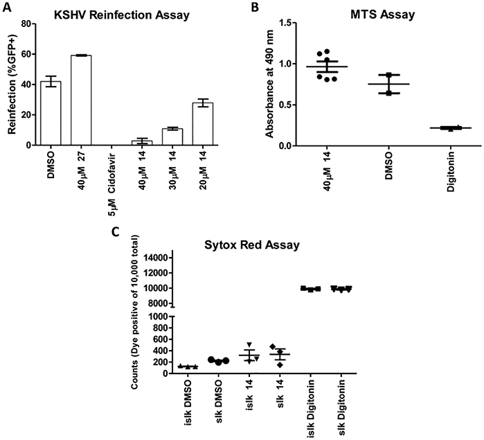
Targeting of cryptic binding sites represents an attractive but underexplored approach to modulating protein function with small molecules. Using the dimeric protease (Pr) from Kaposi's sarcoma-associated herpesvirus (KSHV) as a model system, we sought to dissect a putative allosteric network linking a cryptic site at the dimerization interface to enzyme function. Five cryogenic X-ray structures were solved of the monomeric protease with allosteric inhibitors bound to the dimer interface site. Distinct coordinated movements captured by the allosteric inhibitors were also revealed as alternative states in room-temperature X-ray data and comparative analyses of other dimeric herpesvirus proteases. A two-step mechanism was elucidated through detailed kinetic analyses and suggests an enzyme isomerization model of inhibition. Finally, a representative allosteric inhibitor from this class was shown to be efficacious in a cellular model of viral infectivity. These studies reveal a coordinated dynamic network of atomic communication linking cryptic binding site occupancy and allosteric inactivation of KHSV Pr that can be exploited to target other members of this clinically relevant family of enzymes.


Conflict of interest statement

The authors declare no competing financial interest.

Figures

Figure 1.
Figure 1.
Binding of small molecules to the cryptic binding pocket leads to coordinated rearrangements of distal sites at the protein. (A) The KSHV protease dimer (PDB: 2PBK) is shown with the dimer interface helices in orange. Trp109 is shown in brown and blue and is located behind helix 5. The active-site residues are shown in orange, and the loop regions that adopt distinct conformations in the compound bound monomers are shown in blue. (B) The small-molecule scaffold with variable R-group regions is shown, where Y is either COOH or a tetrazole (Tz). (C) Overlay of the cryogenic co-crystal structures solved in this study (PDB codes: 5UR3, 5UVP, 5UV3, 5UTE, 5UTN). The dynamic loop regions are shown in red, scaled to their B-factors and the compounds are shown in orange. D. One monomer from the dimer structure (2PBK) is overlaid with a monomer from this study. Several loops from the monomeric structures, shown in red, are in distinct conformations from those of the dimeric structure shown in blue. (E) The R1 groups from compounds that are co-crystallized in this study are shown (benzyl (1), 4-F-Benzyl (2), 4-OCH3-Benzyl (3), 3-OCF3-Benzyl (4), tetrahydropyran (14). Note: R2 is cyclohexyl for each of the co-crystallized compounds. (This figure is enlarged in the Supporting Information.)
Figure 2.
Figure 2.
Distinct C-terminal conformations are identified in this study. (A) Compound 1 cryogenic co-crystal structure with the surface representation shown. The orientation of the C-terminal residues forms a well-defined pocket that encapsulates the compound. (B) Compound 14 cryogenic co-crystal structure with the surface representation shown. The orientation of the C-terminal residues leaves the anion exposed to solvent and is in a similar trajectory to that of the dimeric helices. (C) Electron density supporting temperature-dependent conformational differences between structures determined at 100 and 280 K (280 K PDB codes: 5V5D, 5V5E). The leftmost column of panel shows an electron density map and model derived from cryogenic (100 K, compound 4) data, the middle panel shows maps and models derived from room-temperature (280 K, compound 4) data, and the rightmost panel shows overlays of the 100 and 280 K models. The electron density maps are calculated using 2FoFc amplitudes with model phases and are contoured at 2.5σ (yellow) or 1.0σ (blue). A nearly 180° rotation of the φ-angle of Glu194 positions the C-terminus in opposite directions in each of the two structures, leading to a slightly different set of interactions stabilizing the cryptic binding site.
Figure 3.
Figure 3.
Compounds display slow time-dependent inhibition and two-step inhibition. (A) The fractional velocity of the reactions shows that there is a rapid, concentration-dependent inhibition followed by a slow onset to the steady state. The curves are from a two-fold dilution scheme beginning at 25 μM (red) and ending at 1.6 μM (black) of compound 14. (B) Fitting the progress curves for kobs shows a hyperbolic fit for the compounds, supporting a two-step enzyme isomerization mechanism of inhibition. The data shown are for compound 1.
Figure 4.
Figure 4.
Cellular evaluation of a tetrazole compound 14. (A) Compound 14 displays concentration-dependent inhibition in reinfectivity as compared to DMSO. An inactive congener shows no inhibition of re-infectivity. Cidofavir is included as a positive control. (B) Cell viability, as measured by MTS assay shows no significant differences between compound-treated and DMSO control. Digitonin served as a positive control. (C) The Sytox red assay for membrane permeability shows no significant differences between compound-treated and DMSO-treated cells for the iSLK and SLK cells.

References

    1. Human Herpesviruses: Biology, Therapy, and Immunoprophylaxis; Arvin A, Gabriella C-F, Mocarski E, Moore PS, Roizman B, Whitely R, Yamanishi K, Eds.; Cambridge University Press: Cambridge, 2007; https://www.ncbi.nlm.nih.gov/books/NBK47376/. - PubMed
    1. Gable JE; Acker TM; Craik CS Chem. Rev 2014, 114 (22), 11382–11412. - PMC - PubMed
    1. Preston VG; Coates JAV; Rixon FJ J. Virol 1983, 45 (3), 1056–1064. - PMC - PubMed
    1. Shahian T; Lee GM; Lazic A; Arnold LA; Velusamy P; Roels CM; Guy RK; Craik CS Nat. Chem. Biol 2009, 5 (9), 640–646. - PMC - PubMed
    2. Lee GM; Shahian T; Baharuddin A; Gable JE; Craik CSJ J. Mol. Biol 2011, 411 (5), 999–1016. - PMC - PubMed
    1. Cimermancic P; Weinkam P; Rettenmaier TJ; Bichmann L; Keedy DA; Woldeyes RA; Schneidman-Duhovny D; Demerdash ON; Mitchell JC; Wells JA; Fraser JS; Sali AJ J. Mol. Biol 2016, 428 (4), 709–719. - PMC - PubMed

Publication types

MeSH terms