Review
. 2017 Oct 6:33:319-342.
doi: 10.1146/annurev-cellbio-100616-060758. Epub 2017 Jul 31.

Rethinking m6A Readers, Writers, and Erasers

Affiliations
Review

Rethinking m6A Readers, Writers, and Erasers

Kate D Meyer et al. Annu Rev Cell Dev Biol. .

Abstract

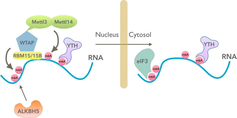
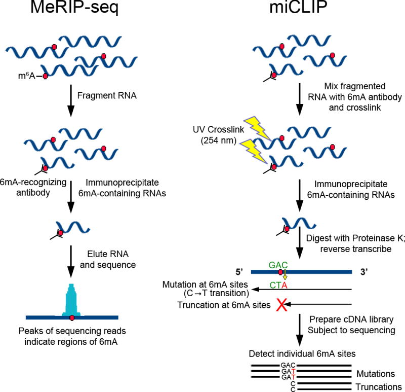
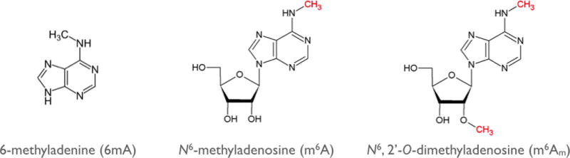
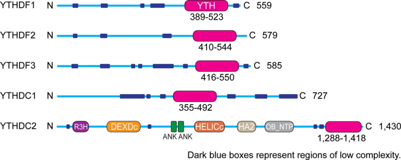
In recent years, m6A has emerged as an abundant and dynamically regulated modification throughout the transcriptome. Recent technological advances have enabled the transcriptome-wide identification of m6A residues, which in turn has provided important insights into the biology and regulation of this pervasive regulatory mark. Also central to our current understanding of m6A are the discovery and characterization of m6A readers, writers, and erasers. Over the last few years, studies into the function of these proteins have led to important discoveries about the regulation and function of m6A. However, during this time our understanding of these proteins has also evolved considerably, sometimes leading to the reversal of early concepts regarding the reading, writing and erasing of m6A. In this review, we summarize recent advances in m6A research, and we highlight how these new findings have reshaped our understanding of how m6A is regulated in the transcriptome.

Keywords: RNA modifications; epitranscriptome; m6A; posttranscriptional control.


Figures

Figure 1
Figure 1
mRNA control by an m6A (N6-methyladenosine) writer complex, an eraser, and readers. The m6A writer complex (RBM15/15B-WTAP-METTL3-METTL14) and the m6A eraser (ALKBH5) are localized primarily in the nucleus. Therefore, any dynamic regulation of m6A in mRNA would occur in the nucleus prior to mRNA export. The m6A “imprint” that is conferred in the nucleus is essentially unchangeable and dictates the mRNA’s fate in the cytoplasm. In the nucleus, m6A can be recognized by m6A readers, the most prominent of which is the YTH protein DC1. In the cytoplasm, m6A can be recognized by the highly similar, and possibly functionally redundant, YTH proteins DF1, DF2, and DF3. Additionally, m6A in 5´UTRs can bind eukaryotic initiation factor 3 (eIF3), which promotes translation independently of the canonical cap-binding protein eIF4E.
Figure 2
Figure 2
Methods for transcriptome-wide mapping of adenosine methylation. Shown here are schematics outlining the general procedures for MeRIP-Seq (a) and miCLIP (b). In both methods, cellular RNA is fragmented and incubated with 6mA (6-methyladenine)-recognizing antibodies. (a) In MeRIP-Seq, methylated RNAs are directly immunoprecipitated using antibody-binding magnetic beads. Methylated RNA is then eluted by either proteinase K digestion or the addition of free m6A (N6-methyladenosine), and RNA fragments are subjected to high-throughput sequencing. The resulting sequencing reads accumulate around sites of 6mA and enable prediction of an ~100–200-nt-wide region of one or more m6A residues. Enriched reads near the 5´ end of RNAs can be due to either m6A or m6Am (N6,2´-O-dimethyladenosine). (b) In miCLIP, 6mA antibodies are crosslinked to methylated RNA prior to immunoprecipitation. Elution with proteinase K leaves a small crosslinked adduct on the methylated RNA, which causes either truncations or mutations immediately adjacent to methylation sites during reverse transcription. The resulting cDNA is then amplified and subjected to next-generation sequencing, and individual m6A or m6Am sites are identified by detecting sites of mutation or truncation in the sequencing reads.
Figure 3
Figure 3
m6A and m6Am are the two 6mA (6-methyladenine)-containing nucleotides in mRNA. 6mA is found in two distinct epitranscriptomic modifications: m6A (N6-methyladenosine) and m6Am (N6,2´-O-dimethyladenosine). The methyl groups that are added enzymatically to alter the function of the nucleotide are shown in red. m6A is found in 5´UTRs, coding sequences, and 3´UTRs. m6Am is found in one place in mRNAs, the first encoded nucleotide, which is adjacent to the m7G (N7-methylguanosine) cap.
Figure 4
Figure 4
Schematic representation of the domain structures of the five human YTH proteins: DF1, DF2, DF3, DC1, and DC2. The YTH domain (magenta) is at the C-terminal region in DF1, DF2, DF3, and DC2 and is located internally in DC1. DC1 has a different domain organization than DC2. DF1, DF2, and DF3 show highly similar sequence and domain structure. The low-complexity (dark blue) and Glu-rich (red) regions are indicated, as are the R3H, DEXDc, ankyrin repeat (ANK), HELICc, HA2, and OB-fold (OB_NTP) domains for DC2. The length of the protein is indicated at the right of each protein schematic.

References

    1. Agarwala SD, Blitzblau HG, Hochwagen A, Fink GR. RNA methylation by the MIS complex regulates a cell fate decision in yeast. PLOS Genet. 2012;8:e1002732. - PMC - PubMed
    1. Aguzzi A, Altmeyer M. Phase separation: linking cellular compartmentalization to disease. Trends Cell Biol. 2016;26:547–58. - PubMed
    1. Alarcon CR, Goodarzi H, Lee H, Liu X, Tavazoie S, Tavazoie SF. HNRNPA2B1 is a mediator of m6A-dependent nuclear RNA processing events. Cell. 2015a;162:1299–308. - PMC - PubMed
    1. Alarcon CR, Lee H, Goodarzi H, Halberg N, Tavazoie SF. N6-Methyladenosine marks primary microRNAs for processing. Nature. 2015b;519:482–85. - PMC - PubMed
    1. Banko JL, Hou L, Poulin F, Sonenberg N, Klann E. Regulation of eukaryotic initiation factor 4E by converging signaling pathways during metabotropic glutamate receptor-dependent long-term depression. J Neurosci. 2006;26:2167–73. - PMC - PubMed