. 2017 Aug;50(8):429-434.
doi: 10.5483/bmbrep.2017.50.8.097.

Transforming growth factor β1 enhances adhesion of endometrial cells to mesothelium by regulating integrin expression

Affiliations

Transforming growth factor β1 enhances adhesion of endometrial cells to mesothelium by regulating integrin expression

Hee-Jung Choi et al. BMB Rep. 2017 Aug.

Abstract

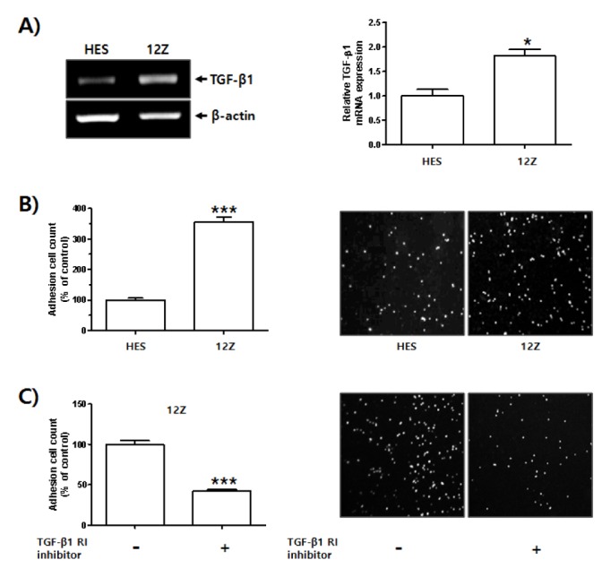
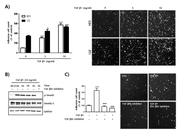
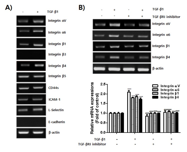
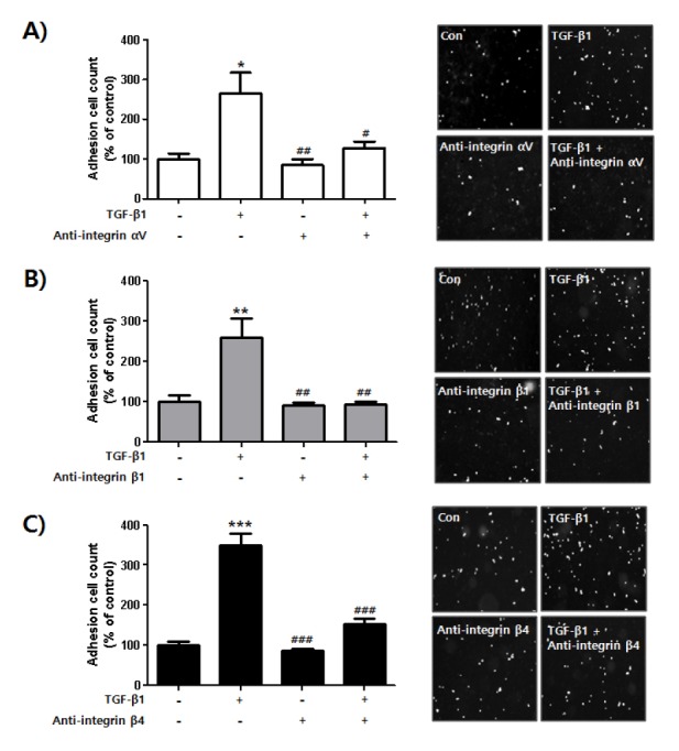
Endometriosis is the abnormal growth of endometrial cells outside the uterus, causing pelvic pain and infertility. Furthermore, adhesion of endometrial tissue fragments to pelvic mesothelium is required for the initial step of endometriosis formation outside uterus. TGF-β1 and adhesion molecules importantly function for adhesion of endometrial tissue fragments to mesothelium outside uterus. However, the function of TGF-β1 on the regulation of adhesion molecule expression for adhesion of endometrial tissue fragments to mesothelium has not been fully elucidated. Interestingly, transforming growth factor β1 (TGF-β1) expression was higher in endometriotic epithelial cells than in normal endometrial cells. The adhesion efficiency of endometriotic epithelial cells to mesothelial cells was also higher than that of normal endometrial cells. Moreover, TGF-β1 directly induced the adhesion of endometrial cells to mesothelial cells through the regulation of integrin of αV, α6, β1, and β4 via the activation of the TGF-β1/TGF-βRI/Smad2 signaling pathway. Conversely, the adhesion of TGF-β1-stimulated endometrial cells to mesothelial cells was clearly reduced following treatment with neutralizing antibodies against specific TGF-β1-mediated integrins αV, β1, and β4 on the endometrial cell membrane. Taken together, these results suggest that TGF-β1 may act to promote the initiation of endometriosis by enhancing integrin-mediated cell-cell adhesion. [BMB Reports 2017; 50(8): 429-434].


Conflict of interest statement

CONFLICTS OF INTEREST

The authors have no conflicting financial interests.

Figures

Fig. 1
Fig. 1
Enhanced TGF-β1 expression in human endometriotic epithelial cells and its function in adhesion of endometrial cells to mesothelial cells. Total RNA was extracted from HES cells and 12Z cells. (A) Levels of TGF-β1 mRNA expression were examined using RT-PCR. β-actin was used as an internal control. Band intensity of TGF-β1 mRNA expression was quantified and normalized to β-actin internal control using densitometry (ImageJ software, NIH). Data obtained from densitometric analyses are shown as bar graph. Data are expressed as fold of control and shown as mean ± SD for three independent experiments (*P < 0.05 in comparison between two groups). Differences between mean values and two groups were evaluated using Student’s t-test and analysis of variance with an unpaired t-test. (B) HES cells (5 × 10⁵ cells) were seeded onto 6-well plate and cultured for 24 h. 12Z cells (3 × 10⁵ cells) were seeded onto 100 π culture dish plate and cultured for 24 h. HES and 12Z cells were labeled with CMFDA for 15 min at 37°C, then washed in 1 × PBS and gently transferred onto a Met-5A cell monolayer. Number of HES and 12Z cells bound to confluent Met-5A cells was manually counted. Four pictures were taken per well and the number of adherent cells was calculated as a percentage of the control cell values and expressed as mean ± SD for three independent experiments (***P < 0.01 in comparison between two groups). Differences between mean values and two groups were evaluated using Student’s t-test and analysis of variance with an unpaired t-test. (C) 12Z cells (3 × 10⁵ cells) were seeded onto 100 π culture dish plate and cultured for 24 h. Medium was replaced and cells were incubated in serum free-medium with or without TGF-βRI inhibitor for 24 h. Cells were then labeled with CMFDA for 15 min at 37°C, then washed in 1 × PBS and gently transferred onto a Met-5A cell monolayer. Number of cells bound to confluent Met-5A cells was manually counted. Four pictures were taken per well and the number of adherent cells was calculated as a percentage of the control cell values and expressed as mean ± SD for three independent experiments (***P < 0.01 in comparison between two groups). Differences between mean values and two groups were evaluated using Student’s t-test and analysis of variance with an unpaired t-test.
Fig. 2
Fig. 2
Increased adhesion of endometrial cells to mesothelial cells by activation of TGF-β1-mediated signaling. (A) HES and 12Z cells were seeded and cultured for 24 h. Medium was replaced and cells were incubated in serum free-medium with or without TGF-β1 for 24 h. Cells were labeled with CMFDA for 15 min at 37°C, then washed in 1 × PBS and gently transferred onto a Met-5A cell monolayer. After gentle shaking at 20 rpm for 20 min at 37°C, Cells were washed three times with 1 × PBS to remove unbound cells. Attached cells were visualized using a fluorescent microscope, and quantified using ImageJ software. The number of cells in 4 randomly chosen areas in each well was used for statistical analysis. The results from 3 independent experiments were calculated as a percentage of the control cell values and presented as mean ± SD. ***P < 0.001 compared to control white bar graph (1st column). #P < 0.05 and ###P < 0.001 compared to control black bar graph (1st column). Differences between mean values of experimental groups were determined using one-way analysis of variance (one-way ANOVA) with a Tukey’s post-hoc test, using GraphPad Prism software. (B) HES cells were seeded and cultured for 24 h. Medium was replaced and cells were incubated in serum free-medium with or without TGF-β1 in the presence or absence of TGF-βRI inhibitor for the indicated times. Phosphorylation levels of Smad2 were analyzed using western blot. GAPDH expression was used as an internal control. (C) HES cells were seeded and cultured for 24 h. Medium was replaced and cells were incubated in serum free-medium with or without TGF-β1 in the presence or absence of TGF-βRI inhibitor for the indicated times. Cells were labeled with CMFDA for 15 min at 37°C, then washed in 1 × PBS and then gently transferred onto a Met-5A cell monolayer. Number of cells bound to confluent Met-5A cells was manually counted. Four pictures were taken per well and the number of adherent cells was calculated as a percentage of the control cell values and shown as mean ± SD for three independent experiments. ***P < 0.001 compared to negative control (1st column). ###P < 0.001 compared to positive control (2nd column). Differences between mean values of experimental groups were determined using one-way ANOVA with a Tukey’s post-hoc test, using GraphPad Prism software.
Fig. 3
Fig. 3
Expressions of integrin αV, α6, β1, and β4 induced by TGF-β1 in endometrial cells. HES cells were seeded and cultured for 24 h. Medium was replaced and cells were incubated in serum free-medium with or without TGF-β1 for 24 h. Total RNA was extracted from the cells. (A) mRNA expression of adhesion molecules was examined using RT-PCR. β-actin was used as an internal control. (B) HES cells were seeded and cultured for 24 h. Medium was replaced and the cells were incubated in serum free-medium with or without TGF-β1 in the presence or absence of TGF-βRI inhibitor for 24 h. Total RNA was extracted from the cells. Expression levels of integrin αV, α6, β1, and β4 were examined using RT-PCR. β-actin was used as an internal control. Band intensity of each integrin mRNA expression was quantified and normalized to β-actin internal control using densitometry. Data obtained from densitometric analyses are shown as bar graph. Data are expressed as fold of control and are shown as mean ± SD for three independent experiments ***P < 0.001 compared to each negative control (1st column). ###P < 0.001 compared to each positive control (2nd column). Differences between mean values of experimental groups were determined using a one-way ANOVA with a Tukey’s post-hoc test, using GraphPad Prism software.
Fig. 4
Fig. 4
Blocking adhesion of TGF-β1-stimulated endometrial cells to mesothelial cells using integrin αV, β1, and β4 neutralizing antibodies. HES cells were seeded and cultured for 24 h. Medium was replaced and cells were incubated in serum free-medium with or without TGF-β1 in the presence or absence of integrin (A) αV, (B) β1, or (C) β4 antibodies for 24 h. Cells were labeled with CMFDA for 15 min at 37°C, then washed in 1 × PBS and gently transferred onto a Met-5A cell monolayer. Number of cells bound to confluent Met-5A cells was manually counted. Four pictures were taken per well and the number of adherent cells was calculated as a percentage of the control cell values and shown as mean ± SD for three independent experiments. *P < 0.05, **P < 0.01, ***P < 0.001 compared to each negative control (1st column of each graph). #P < 0.05, ##P < 0.01, ###P < 0.001 compared to each positive control (2nd column of each graph). Differences between mean values of experimental groups were determined using a one-way ANOVA with a Tukey’s post-hoc test, using GraphPad Prism software.

Similar articles

Cited by

References

    1. Ping S, Ma C, Liu P, et al. Molecular mechanisms underlying endometriosis pathogenesis revealed by bioinformatics analysis of microarray data. Arch Gynecol Obstet. 2016;293:797–804. doi: 10.1007/s00404-015-3875-y. - DOI - PubMed
    1. Nothnick W, Alali Z. Recent advances in the understanding of endometriosis: the role of inflammatory mediators in disease pathogenesis and treatment. F1000Res. 2016:5. - PMC - PubMed
    1. Bedir R, Sehitoglu I, Balik G, et al. The role of the adhesion molecule Nectin-4 in the pathogenesis of endometriosis. Clin Exp Obstet Gynecol. 2016;43:463–466. - PubMed
    1. Schutt AK, Atkins KA, Slack-Davis JK, Stovall DW. VCAM-1 on peritoneum and alpha4beta1 integrin in endometrium and their implications in endometriosis. Int J Gynecol Pathol. 2015;34:85–89. doi: 10.1097/PGP.0000000000000118. - DOI - PubMed
    1. Sundqvist J, Andersson KL, Scarselli G, Gemzell-Danielsson K, Lalitkumar PG. Expression of adhesion, attachment and invasion markers in eutopic and ectopic endometrium: a link to the aetiology of endometriosis. Hum Reprod. 2012;27:2737–2746. doi: 10.1093/humrep/des220. - DOI - PubMed