. 2017 Sep 19;199(20):e00281-17.
doi: 10.1128/JB.00281-17. Print 2017 Oct 15.

TetR Family Transcriptional Regulator PccD Negatively Controls Propionyl Coenzyme A Assimilation in Saccharopolyspora erythraea

Affiliations

TetR Family Transcriptional Regulator PccD Negatively Controls Propionyl Coenzyme A Assimilation in Saccharopolyspora erythraea

Zhen Xu et al. J Bacteriol. .

Abstract

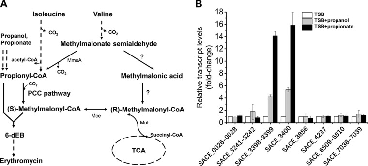
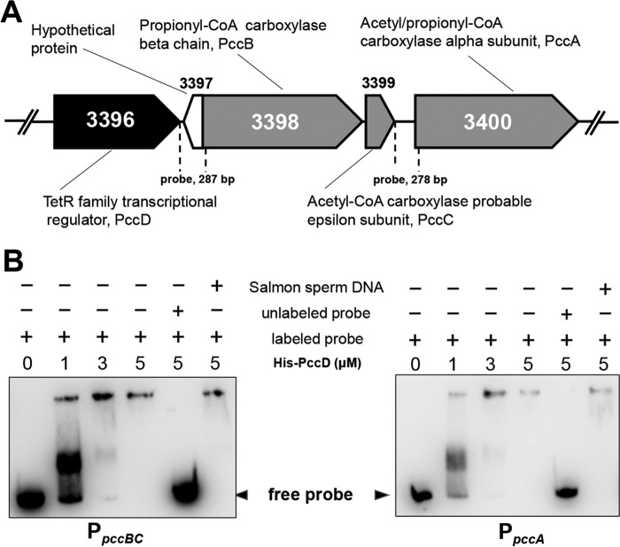
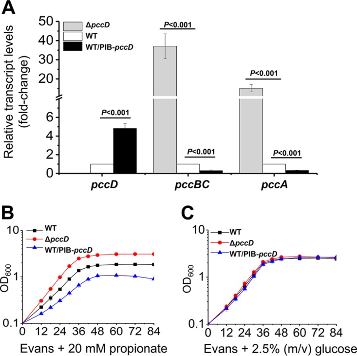
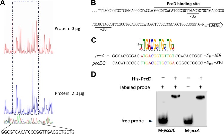
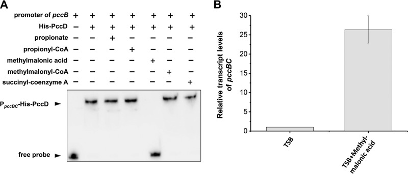
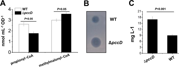
Propanol stimulates erythromycin biosynthesis by increasing the supply of propionyl coenzyme A (propionyl-CoA), a starter unit of erythromycin production in Saccharopolyspora erythraea Propionyl-CoA is assimilated via propionyl-CoA carboxylase to methylmalonyl-CoA, an extender unit of erythromycin. We found that the addition of n-propanol or propionate caused a 4- to 16-fold increase in the transcriptional levels of the SACE_3398-3400 locus encoding propionyl-CoA carboxylase, a key enzyme in propionate metabolism. The regulator PccD was proved to be directly involved in the transcription regulation of the SACE_3398-3400 locus by EMSA and DNase I footprint analysis. The transcriptional levels of SACE_3398-3400 were upregulated 15- to 37-fold in the pccD gene deletion strain (ΔpccD) and downregulated 3-fold in the pccD overexpression strain (WT/pIB-pccD), indicating that PccD was a negative transcriptional regulator of SACE_3398-3400. The ΔpccD strain has a higher growth rate than that of the wild-type strain (WT) on Evans medium with propionate as the sole carbon source, whereas the growth of the WT/pIB-pccD strain was repressed. As a possible metabolite of propionate metabolism, methylmalonic acid was identified as an effector molecule of PccD and repressed its regulatory activity. A higher level of erythromycin in the ΔpccD strain was observed compared with that in the wild-type strain. Our study reveals a regulatory mechanism in propionate metabolism and suggests new possibilities for designing metabolic engineering to increase erythromycin yield.IMPORTANCE Our work has identified the novel regulator PccD that controls the expression of the gene for propionyl-CoA carboxylase, a key enzyme in propionyl-CoA assimilation in S. erythraea PccD represses the generation of methylmalonyl-CoA through carboxylation of propionyl-CoA and reveals an effect on biosynthesis of erythromycin. This finding provides novel insight into propionyl-CoA assimilation, and extends our understanding of the regulatory mechanisms underlying the biosynthesis of erythromycin.

Keywords: erythromycin biosynthesis; propionate metabolism; propionyl-CoA assimilation; transcriptional regulation.


Figures

FIG 1
FIG 1
pccBC (SACE_3398–3399) and pccA (SACE_3400) operons are induced by propanol and propionate in S. erythraea. (A) In S. erythraea, branched-chain amino acids (BCAA) are a source of propionyl-CoA and methylmalonyl-CoA, key precursors in erythromycin biosynthesis. Valine and isoleucine were metabolized to propionyl-CoA through the degradation pathways. Propanol and propionate are two important external sources of propionyl-CoA, especially in the production of erythromycin. The metabolism of propionyl-CoA to (2S)-methylmalonyl–CoA is via the PCC pathway in S. erythraea, which plays an important role in propionyl-CoA assimilation when n-propanol or valine is added for high production of erythromycin. Enzymes that transform methylmalonate-semialdehyde to (R)-methylmalonyl–CoA (indicated by question marks) have not yet been investigated in S. erythraea. (B) Total RNA of S. erythraea NRRL2338 was extracted after 36 h of growth (see Fig. S3 in the supplemental material) in TSB medium, TSB supplied with 1% (vol/vol) n-propanol, and TSB supplied with 20 mM propionate. Relative transcript levels were normalized to the 16S rRNA. The transcription value of each gene in S. erythraea in TSB medium was arbitrarily normalized to 1. Error bars represent the standard deviations from three biological replicates. MmsA, methylmalonate-semialdehyde dehydrogenase (acylating); Mut, methylmalonyl-CoA mutase; Mce, methylmalonyl-CoA epimerase.
FIG 2
FIG 2
PccD (SACE_3396) binds to the promoter regions of pccBC and pccA. (A) Genetic organization of the pccD gene in the S. erythraea genome. pccB (SACE_3398), pccC (SACE_3399), and pccA (SACE_3400) encode a propionyl-CoA carboxylase β-chain, acetyl-CoA carboxylase probable ε-subunit, and acetyl/propionyl-CoA carboxylase α-subunit, respectively. The numbers between dashed lines show the lengths of the promoter regions of pccBC and pccA, respectively, used in the gel shift experiment shown in panel B. (B) EMSA of His-PccD protein with promoter regions (separated by dashed lines shown in panel A) of pccBC or pccA. The biotin-labeled DNA probe (about 15 ng, reaction system 10 μl) was incubated with a protein concentration gradient (0, 1, 3, and 5 μM). An unlabeled specific probe (200-fold) or nonspecific competitor DNA (200-fold, sonicated salmon sperm DNA) was used as control. The free probes that did not bind with protein are shown by arrowheads.
FIG 3
FIG 3
PccD is a repressor of pccBC and pccA operons. (A) qPCR analysis of the relative transcription levels of pccD, pccBC, and pccA genes between the WT, ΔpccD, and WT/PIB-pccD strains. Total RNA of these strains was extracted after 36 h of growth in TSB medium (see Fig. S5A in the supplemental material). The expression levels of genes in the WT strain were arbitrarily set to 1.0. Error bars indicate standard deviation from three independent experiments. (B and C) Growth curves of S. erythraea strains WT, ΔpccD, and WT/PIB-pccD grown on Evans medium supplemented with 20 mM sodium propionate (B) or 2.5% glucose (C) as the sole carbon source. Error bars indicate standard deviation from three biological replicates.
FIG 4
FIG 4
Identification of a PccD-binding site in pccBC and pccA operons. (A) Electropherograms of a DNase I digest of pccBC promoter probe incubated without (top) or with (bottom) 2.0 μg of His-PccD. The nucleotide sequence protected by His-PccD is indicated below. (B) Protected sequence stretches in the upstream regions of pccBC. Black lines indicate the regions of DNase I protection. The −35 and −10 sites were predicted by the Softberry web tool. (C) Analysis of the PccD-binding motif of pccA using MEME. The standard code of the WebLogo server is shown at the top. (D) Verification of PccD-binding motif by EMSA. Synthetic probes M-pccBC and M-pccA containing PccD-binding site in promoters of pccBC and pccA operons, respectively. The DNA probe was incubated with a protein concentration of 5 μM.
FIG 5
FIG 5
The metabolite methylmalonic acid is an inhibitor of PccD. (A) EMSA of His-PccD protein binding to the upstream promoter regions of pccBC. The biotin-labeled DNA probe (about 15 ng, reaction system 10 μl) was incubated with 5 μM His-PccD. Propionate, propionyl-CoA, methylmalonic acid, methylmalonyl-CoA, or succinyl-CoA was added as a cofactor to a final concentration of 1.0 mM. (B) Total RNA of S. erythraea NRRL2338 was extracted after 36 h of growth in TSB medium or TSB supplied with 10 mM methylmalonic acid. Relative transcript levels were normalized to the 16S rRNA. The transcription value of pccBC in S. erythraea WT cultivated in TSB medium was arbitrarily normalized to 1. Error bars represent the standard deviations from three biological replicates.
FIG 6
FIG 6
Apparent increase of erythromycin production in the pccD deletion strain compared to wild type. (A) Intracellular propionyl-CoA and methylmalonyl-CoA concentration of S. erythraea WT and ΔpccD strains grown in TSB medium with 1% (vol/vol) n-propanol. Cells were harvested at 36 h (see Fig. S5B in the supplemental material). (B) Inhibition tests of S. erythraea WT and ΔpccD fermentation broths, collected after culturing for 84 h, against Bacillus subtilis. (C) Erythromycin concentration of S. erythraea WT and ΔpccD strains grown in TSB medium with 1% (vol/vol) n-propanol. Supernatants were collected after culture for 84 h. A turbidimetric method for microbiological assay of antibiotics was used to quantify the erythromycin levels as described in Materials and Methods. Three independent experiments were performed to calculate standard deviation.

References

    1. Staunton J, Weissman KJ. 2001. Polyketide biosynthesis: a millennium review. Nat Prod Rep 18:380–416. doi:10.1039/a909079g. - DOI - PubMed
    1. Li L, Zhao Y, Ruan L, Yang S, Ge M, Jiang W, Lu Y. 2015. A stepwise increase in pristinamycin II biosynthesis by Streptomyces pristinaespiralis through combinatorial metabolic engineering. Metab Eng 29:12–25. doi:10.1016/j.ymben.2015.02.001. - DOI - PubMed
    1. Wang W, Tian J, Li L, Ge M, Zhu H, Zheng G, Huang H, Ruan L, Jiang W, Lu Y. 2015. Identification of two novel regulatory genes involved in pristinamycin biosynthesis and elucidation of the mechanism for AtrA-p-mediated regulation in Streptomyces pristinaespiralis. Appl Microbiol Biotechnol 99:7151–7164. doi:10.1007/s00253-015-6638-6. - DOI - PubMed
    1. Zhao Y, Feng R, Zheng G, Tian J, Ruan L, Ge M, Jiang W, Lu Y. 2015. Involvement of the TetR-type regulator PaaR in the regulation of pristinamycin I biosynthesis through an effect on precursor supply in Streptomyces pristinaespiralis. J Bacteriol 197:2062–2071. doi:10.1128/JB.00045-15. - DOI - PMC - PubMed
    1. Staunton J, Wilkinson B. 1997. Biosynthesis of erythromycin and rapamycin. Chem Rev 97:2611–2630. doi:10.1021/cr9600316. - DOI - PubMed

Publication types

LinkOut - more resources