. 2017 Aug 15;114(33):8806-8811.
doi: 10.1073/pnas.1705084114. Epub 2017 Jul 31.

Presumptive TRP channel CED-11 promotes cell volume decrease and facilitates degradation of apoptotic cells in Caenorhabditis elegans

Affiliations

Presumptive TRP channel CED-11 promotes cell volume decrease and facilitates degradation of apoptotic cells in Caenorhabditis elegans

Kaitlin Driscoll et al. Proc Natl Acad Sci U S A. .

Abstract

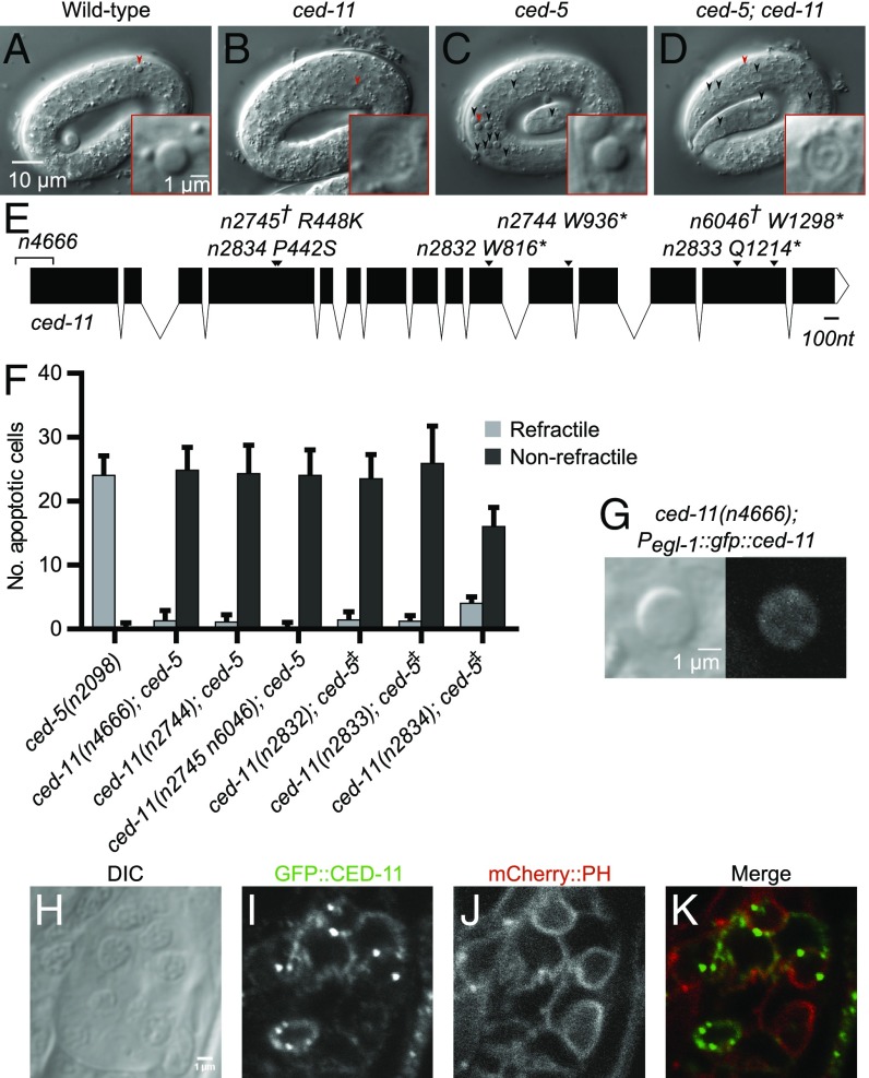
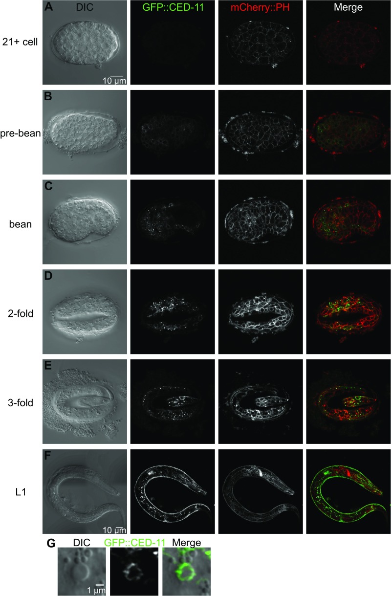
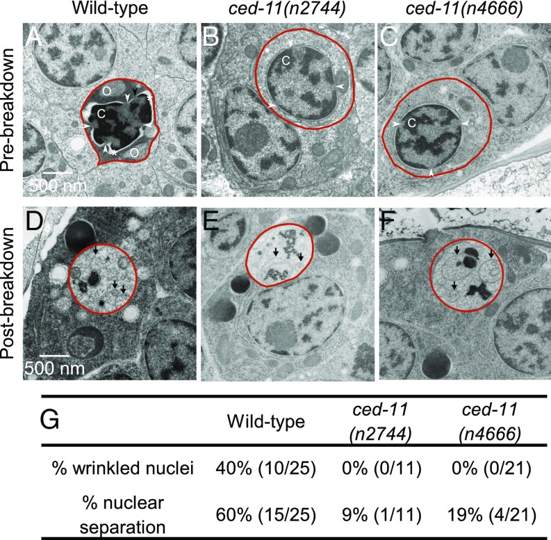
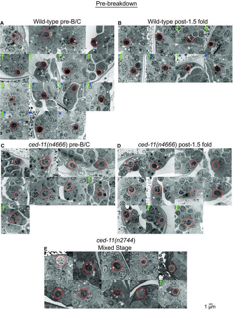
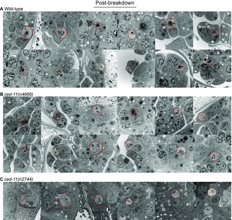
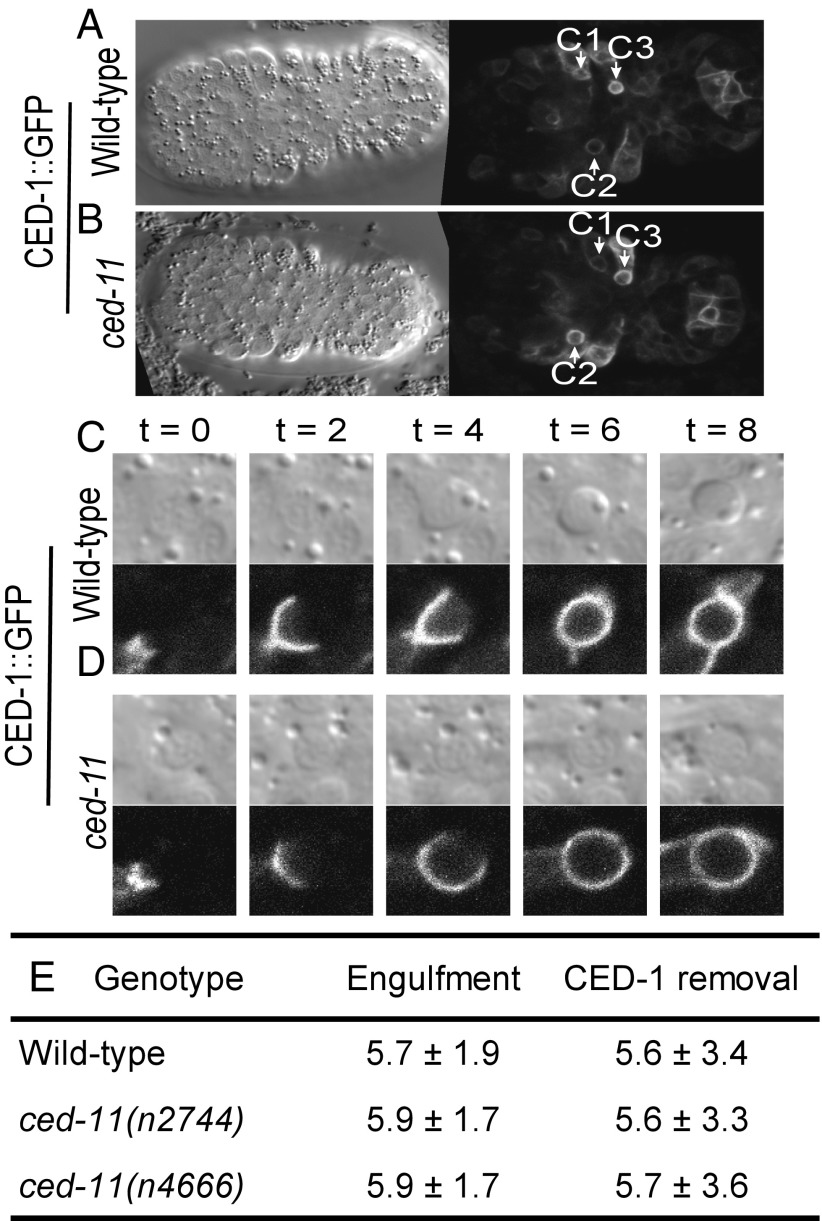
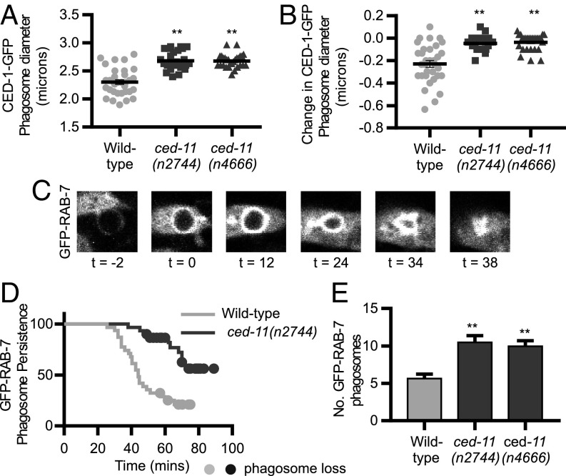
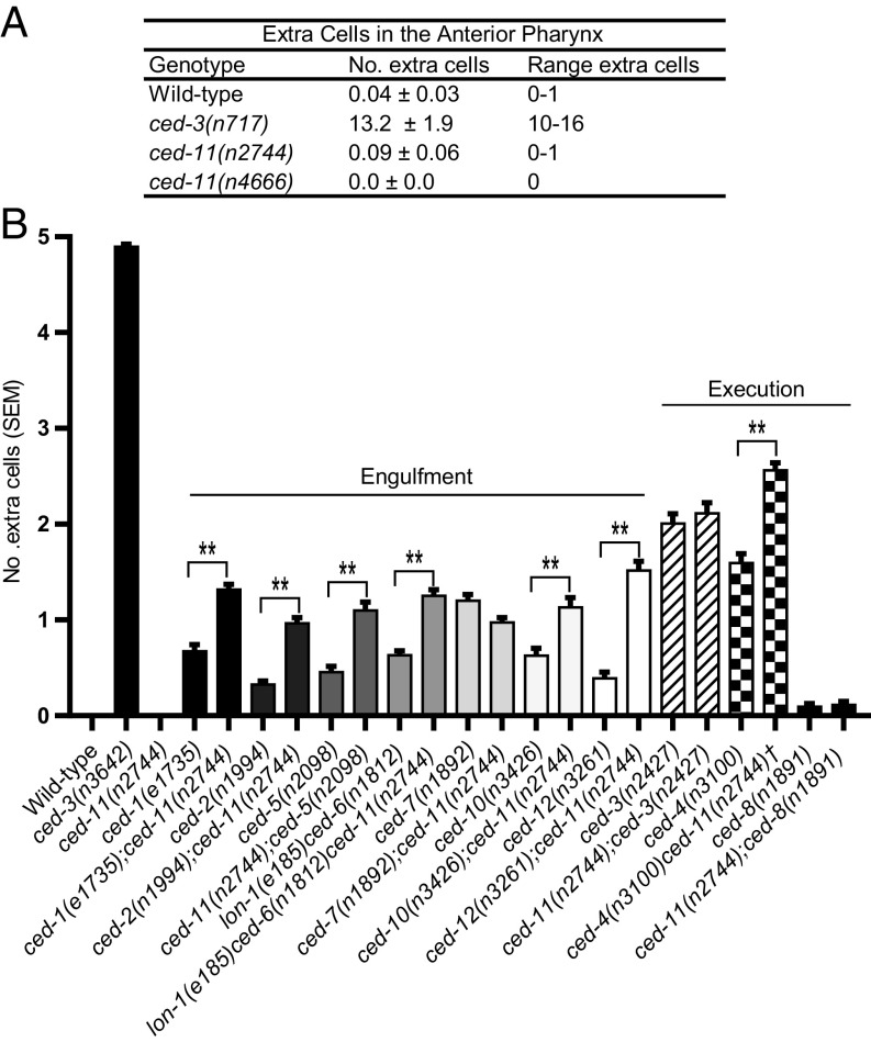
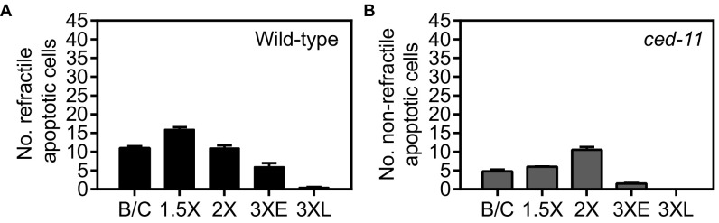
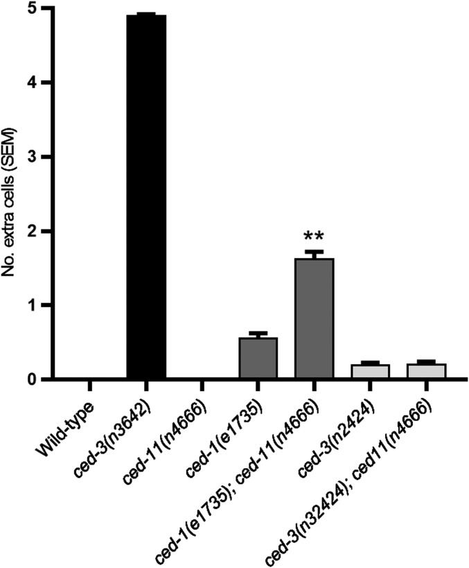
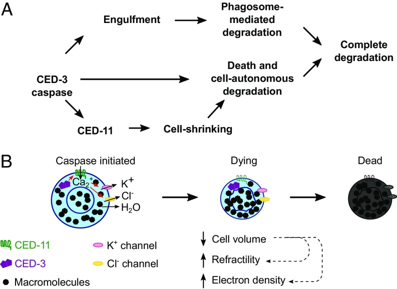
Apoptotic cells undergo a series of morphological changes. These changes are dependent on caspase cleavage of downstream targets, but which targets are significant and how they facilitate the death process are not well understood. In Caenorhabditis elegans an increase in the refractility of the dying cell is a hallmark morphological change that is caspase dependent. We identify a presumptive transient receptor potential (TRP) cation channel, CED-11, that acts in the dying cell to promote the increase in apoptotic cell refractility. CED-11 is required for multiple other morphological changes during apoptosis, including an increase in electron density as visualized by electron microscopy and a decrease in cell volume. In ced-11 mutants, the degradation of apoptotic cells is delayed. Mutation of ced-11 does not cause an increase in cell survival but can enhance cell survival in other cell-death mutants, indicating that ced-11 facilitates the death process. In short, ced-11 acts downstream of caspase activation to promote the shrinkage, death, and degradation of apoptotic cells.

Keywords: C. elegans; TRP channel; apoptosis; apoptotic volume decrease; cell volume.


Conflict of interest statement

The authors declare no conflict of interest.

Figures

Fig. 1.
Fig. 1.
ced-11 is required for the increase in apoptotic cell refractility. (AD) DIC images of threefold-stage embryos. Arrowheads indicate apoptotic cells that are shown in the Insets. (A) Wild-type embryo with refractile apoptotic cell. (B) ced-11(n2744) embryo with nonrefractile apoptotic cell. (C) ced-5(n2098) embryo with accumulated refractile apoptotic cells. (D) ced-11(n2744); ced-5(n2098) embryo with accumulated nonrefractile apoptotic cells. (E) Diagram of the ced-11 gene. Arrows indicate the location of the ced-11 allele. The asterisk indicates the stop codon. The dagger indicates two ced-11 mutations isolated in the same strain. The bracket indicates a deletion. (F) Average numbers of refractile and nonrefractile apoptotic cells in the heads of threefold-stage embryos of the indicated genotypes. ced-5(n2098) was in all strains. The double-dagger indicates that the strain also includes sem-4(n1378). Error bars indicate SD. (n > 20). (G) DIC and GFP images of a refractile apoptotic cell that expresses GFP::CED-11 in a ced-11(n4666) embryo [ced-11(n4666); nEx2544[Pegl-1::gfp::ced-11]. (HK) Head of an L1 larva showing expression of nIs790[Pced-11::gfp::ced-11]; nEx2344[Pevl-20::mCherry::ph. (H) DIC. (I) GFP::CED-11 expression in multiple cells. (J) Plasma membrane marker mCherry::PH. (K) Merged image shows colocalization of GFP::CED-11 and mCherry::PH.
Fig. S1.
Fig. S1.
Sequence alignment of C. elegans CED-11, human TRPM1 (NP_001238949), and human TRPM3 (NP_066003). Black-shaded boxes indicate identical residues, and gray-shaded boxes indicate similar residues.
Fig. S2.
Fig. S2.
Pced-11::gfp::ced-11 expression. (AF) GFP::CED-11 expression pattern in embryos at the indicated stages (AE) and in a larva (F). mCherry::PH localizes to the plasma membrane. mCherry images were taken with different gains to enhance visualization. GFP images in A–E were taken with the same gain to allow comparison between embryos. (Scale bar: 10 µm for all embryos.) The GFP gain in F is higher than in the images of embryos to enhance visualization. (G) DIC image of an apoptotic cell with GFP::CED-11 expression.
Fig. 2.
Fig. 2.
Apoptotic cells in ced-11 animals have altered ultrastructure. (AF) Electron micrographs of engulfed apoptotic cells (outlined in red). (A) A prebreakdown apoptotic cell in a wild-type embryo stained darkly and had a wrinkled nucleus and separation of the nuclear membrane (indicated by the asterisk). The arrowheads indicate the nuclear pore junction. C, condensed chromatin; O, bloated organelle. (B and C) Prebreakdown apoptotic cells in ced-11(n2744) (B) and ced-11(n4666) (C) embryos did not stain darkly and were not distinct from the surrounding living cells. (DF) Apoptotic corpses postbreakdown in wild-type (D), ced-11(n2744) (E), and ced-11(n4666) (F) embryos contained membranous whorls and did not stain darkly. Arrows indicate a membranous whorl. (G) Percent of prebreakdown apoptotic cells in wild-type and ced-11 embryos with wrinkled nuclei and nuclear separation.
Fig. S3.
Fig. S3.
Electron micrographs of prebreakdown apoptotic cells in embryos of indicated genotypes and stages. (A) Wild-type pre-bean and comma (B/C) embryos. (B) Wild-type 1.5-fold–stage to threefold-stage embryos. (C) ced-11(n4666) pre-B/C embryos. (D) ced-11(n4666) 1.5-fold–stage to threefold-stage embryos. (E) ced-11(n2744) mixed-stage embryos. Asterisks mark apoptotic cells with a wrinkled nucleus. Daggers mark an apoptotic cell with separation of the nucleus and cytoplasm. Red circles mark apoptotic cells.
Fig. S4.
Fig. S4.
Electron micrographs of postbreakdown corpses in wild-type (A), ced-11(n4666) (B), and ced-11(n2744) (C) embryos. Red circles mark apoptotic cells.
Fig. 3.
Fig. 3.
Apoptotic cells in ced-11 embryos are engulfed normally. (A and B) DIC (single focal plane, so not all corpses can be seen) and CED-1::GFP (merge of multiple focal planes) images of wild-type (A) and ced-11 (B) embryos at the onset of ventral enclosure when apoptotic cells C1, C2, and C3 have been engulfed. All strains contain enIs7 (Pced-1::ced-1::gfp). (C and D) CED-1::GFP clustering around apoptotic cells as they are engulfed in wild-type (C) and ced-11 (D) embryos with accompanying DIC images. Images were recorded every 2 min; time 0 is determined by the time of the image taken before CED-1::GFP began to encircle the apoptotic cell. Cells are engulfed at 6 min. (E) Average times for CED-1::GFP accumulation (engulfment) and removal (phagosome maturation) in wild-type and ced-11 embryos. At least nine embryos and 26 apoptotic cells were analyzed for each genotype. Mean time is shown in minutes ± SD.
Fig. 4.
Fig. 4.
ced-11 is required for the decrease in cell volume and efficient degradation of apoptotic cells. (A) Diameter of apoptotic cells C1, C2, and C3 in wild-type, ced-11(n2744), and ced-11(n4666) embryos at the time of CED-1::GFP engulfment. We measured the diameter at the most central Z section of each apoptotic cell using CED-1::GFP. Each dot, square, or triangle represents one apoptotic cell. Bars indicate the mean ± SD. At least nine embryos and 26 apoptotic cells were measured for each genotype. **P < 0.0001, one-way ANOVA with Dennett’s correction. (B) Change in the diameter of the apoptotic cells in A, between the time of engulfment and 2 min after engulfment. Each dot, square, or triangle represents one apoptotic cell. Bars indicate the mean ± SD. At least nine embryos and 26 apoptotic cells were measured for each genotype. **P < 0.0001, one-way ANOVA with Dennett's correction. (C) Apoptotic cell degradation was measured using GRP::RAB-7. Time 0 is the onset of GFP::RAB-7 recruitment to the apoptotic cell containing the phagosome. The survival endpoint is when the GFP::RAB-7 apoptotic cell has shrunk and disappeared. (D) Kaplan–Meier survival curve of apoptotic cells (C1, C2, and C3) in wild-type and ced-11(n2744) embryos; GFP-RAB-7 was used to measure degradation times. Apoptotic cells were occasionally lost from the field of view around 50 min, when embryos started moving; circles indicate apoptotic cells that were lost to view before degradation was complete. Ten embryos and 30 apoptotic cells were evaluated for each genotype. P < 0.0001, Gehan–Breslow–Wilcoxon test. (E) Number of GFP::RAB-7+ phagosome-engulfed apoptotic cells in fourfold-stage embryos of the indicated genotypes. Bars indicate mean and SEM; n > 50. **P < 0.0001, one-way ANOVA with Dennett’s correction.
Fig. 5.
Fig. 5.
Mutation of ced-11 does not cause cell survival on its own but can enhance cell survival in other cell-death mutants. (A) Average number of extra cells present in the anterior pharynges of L3–L4 hermaphrodites. Data are shown as mean ± SD; n > 20). (B) Average number of extra cells in the ventral cords of L4 hermaphrodites. Error bars indicate SEM; n > 100. All strains contain nIs106. The ced-11 allele used was ced-11(n2744). The dagger indicates the presence of dpy-18. **P < 0.001, one-way ANOVA with Sidak’s multiple comparisons test.
Fig. S5.
Fig. S5.
Average number of refractile and nonrefractile apoptotic cells identified by DIC in embryos at different stages. (A) Wild-type. (B) ced-11(n2744). Stages: B/C, bean and comma; 1.5×, 1.5-fold–stage embryos; 2×, twofold-stage embryos; 3×E, early threefold-stage embryos; 3×L, late threefold-stage embryos with a well-developed pharynx and grinder. Error bars indicate SEM; n > 20.
Fig. S6.
Fig. S6.
Average number of extra cells in the ventral cords of L4 hermaphrodites. All strains contain nIs106. Error bars indicate SEM; n > 100. **P < 0.0001, unpaired Student’s t test.
Fig. 6.
Fig. 6.
Model for CED-11 function in apoptosis. CED-11 acts downstream of CED-3 to promote cell-autonomous death and degradation in parallel with engulfment and phagosome-mediated degradation. CED-11 is directly or indirectly activated by the CED-3 caspase. CED-11 activation results in a decrease in cell volume, driving an increase in macromolecule density and leading to the observed increases in cellular refractility and electron density. CED-11–mediated changes in the apoptotic cell promote its death and degradation. (A) Pathway. (B) Schematic.

References

    1. Kerr JFR, Wyllie AH, Currie AR. Apoptosis: A basic biological phenomenon with wide-ranging implications in tissue kinetics. Br J Cancer. 1972;26:239–257. - PMC - PubMed
    1. Wyllie AH. Glucocorticoid-induced thymocyte apoptosis is associated with endogenous endonuclease activation. Nature. 1980;284:555–556. - PubMed
    1. Igney FH, Krammer PH. Death and anti-death: Tumour resistance to apoptosis. Nat Rev Cancer. 2002;2:277–288. - PubMed
    1. Poon IKH, Lucas CD, Rossi AG, Ravichandran KS. Apoptotic cell clearance: Basic biology and therapeutic potential. Nat Rev Immunol. 2014;14:166–180. - PMC - PubMed
    1. Lüthi AU, Martin SJ. The CASBAH: A searchable database of caspase substrates. Cell Death Differ. 2007;14:641–650. - PubMed

Publication types

MeSH terms

Substances

LinkOut - more resources