Comparative Study
. 2017 Jul 31;7(1):6895.
doi: 10.1038/s41598-017-07255-z.

Improving cartilage phenotype from differentiated pericytes in tunable peptide hydrogels

Affiliations
Comparative Study

Improving cartilage phenotype from differentiated pericytes in tunable peptide hydrogels

Enateri V Alakpa et al. Sci Rep. .

Abstract

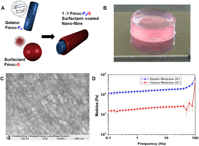
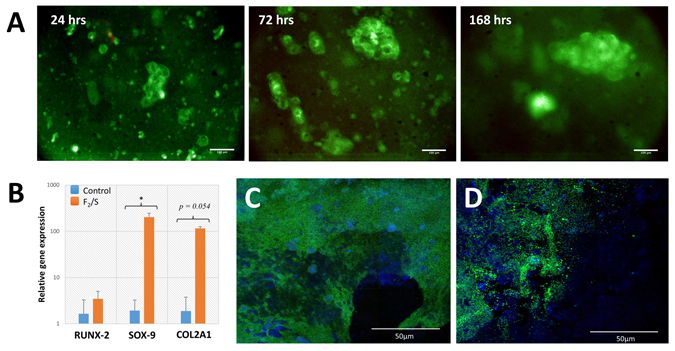
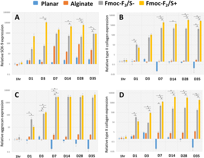
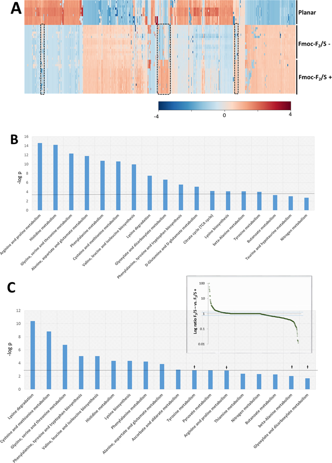
Differentiation of stem cells to chondrocytes in vitro usually results in a heterogeneous phenotype. This is evident in the often detected over expression of type X collagen which, in hyaline cartilage structure is not characteristic of the mid-zone but of the deep-zone ossifying tissue. Methods to better match cartilage developed in vitro to characteristic in vivo features are therefore highly desirable in regenerative medicine. This study compares phenotype characteristics between pericytes, obtained from human adipose tissue, differentiated using diphenylalanine/serine (F2/S) peptide hydrogels with the more widely used chemical induced method for chondrogenesis. Significantly higher levels of type II collagen were noted when pericytes undergo chondrogenesis in the hydrogel in the absence of induction media. There is also a balanced expression of collagen relative to aggrecan production, a feature which was biased toward collagen production when cells were cultured with induction media. Lastly, metabolic profiles of each system show considerable overlap between both differentiation methods but subtle differences which potentially give rise to their resultant phenotype can be ascertained. The study highlights how material and chemical alterations in the cellular microenvironment have wide ranging effects on resultant tissue type.


Conflict of interest statement

RVU is CSO of Biogelx, a spinout company that markets the gels developed and used in this paper.

Figures

Figure 1
Figure 1
Self-assembly of two-component gelators. (A) Schematic presentations of the building blocks, gelator Fmoc-F2, surfactant Fmoc-S and surfactant coated nano fiber Fmoc-F2/S. (B) Macroscopic image for gel in culture media. (C) TEM image of hydrogel showing fibrous morphology. (D) Oscillatory rheology of the gels showing elastic moduli of 15.5 kPa.
Figure 2
Figure 2
(A) Human adipose derived pericyte cultured within Fmoc-F2/S hydrogels. Cells were encapsulated in F2/S hydrogels and maintained in unsupplemented basal media for up to 1 week. Cells were checked for viability by fluorescence detection of Syto 10 (green) for live cells and ethidium homodimer-1 (red) for dead cells after 1, 3 and 7 days. (B) QRT-PCR analysis for gene expression of pericyte cells cultured within 15.5 kPa Fmoc-F2/S hydrogels. Cells were assessed for the production of chondrogenic biomarkers RUNX-2, SOX-9 & type II collagen (COL2A1) after one week in culture. (C & D) Confocal microscopy images of immunofluorescently stained F2/S hydrogels cultured with pericytes for 28 days. Pericytes were checked for chondrogenic development by staining for aggrecan production (C) and type II collagen (D) both ascertained through green fluorescence. Cell populations are indicated by staining the cell nucleus with DAPI (blue). The images are mosaics of a 3 × 3 tile scan, each acquired from random positions of the hydrogel. Scale bar in A is 100 µm, in C & D is 50 µm. In B, Error bars denote the standard error where p < 0.05 as calculated using unpaired student t-test.
Figure 3
Figure 3
Quantitative PCR analysis assessing chondrocyte development of pericytes encapsulated in Fmoc-F2/S hydrogels with (+), without (−) chondrogenic induction and in alginate hydrogels (ALG) also cultured with chondrogenic induction media. Cells were assessed for gene expression of the cartilage biomarkers SOX-9 (A), type II collagen (B), aggrecan (C) and type X collagen (D) up to 35 days in culture. Expression levels of all four biomarker were observed as elevated for all culture systems with the highest expression levels noted for pericytes cultured in Fmoc-F2/S hydrogels. Gene expression was compared against pericytes cultured on glass cover slips (undifferentiated on planar substrate) as a negative control. Error bars are standard error of the mean; * indicate significant difference between groups as determined by one-way ANOVA followed by Bonferroni post hoc test where p < 0.05; n = 4.
Figure 4
Figure 4
Comparison of chondrocyte expression of pericyte cells differentiated over a 5 week period using 15.5 kPa Fmoc-F2/S hydrogels in the presence (+) and absence (−) of chondrogenic induction media. Cells were also differentiated using alginate hydrogels and chondrogenic induction media. (A) Expression of type II collagen relative to aggrecan showed higher type II collagen transcription for cells cultured in Fmoc-F2/S + hydrogels. (B) Expression of type II collagen relative to type X collagen. Gene expression was compared against pericytes cultured on glass cover slips (undifferentiated on planar substrate) as a negative control. Error bars are standard error of the mean; * indicate significant difference between groups as determined by one-way ANOVA followed by Bonferroni post hoc test where p < 0.05; n = 4.
Figure 5
Figure 5
Analysis of metabolite masses detected from pericyte cell extracts undergoing chondrogenesis (n ≥ 12). (A) Hierarchical cluster analysis of metabolic MS masses detected using LC-MS for pericytes cultured on planar substrates (undifferentiated) and cultured in Fmoc-F2/S in the presence or absence of chondrogenic induction media 35 days. (B) Metabolites were mapped to metabolic pathways to ascertain which cell processes are significantly changed from the control. (C) Comparisons between Fmoc-F2/S− and Fmoc-F2/S+ isolate metabolite masses that differ between the two by more than two fold (inset) giving insight into processes that lead to the altered chondrocyte phenotype between Fmoc-F2/S− and Fmoc-F2/S+ Pathways marked with Ɨ were observed to be significantly different from undifferentiated cells but not between Fmoc-F2/S− and Fmoc-F2/S+ suggesting influence during chondrogenesis but do not contribute to the observed differences in chondrocyte phenotype.

References

    1. Muraglia A, et al. Formation of a chondro-osseous rudiment in micromass cultures of human bone-marrow stromal cells. Journal of Cell Science. 2003;116:2949–2955. doi: 10.1242/jcs.00527. - DOI - PubMed
    1. Sekiya I, Vuoristo JT, Larson BL, Prockop DJ. In vitro cartilage formation by human adult stem cells from bone marrow stroma defines the sequence of cellular and molecular events during chondrogenesis. Proceedings of the National Academy of Sciences of the United States of America. 2002;99:4397–4402. doi: 10.1073/pnas.052716199. - DOI - PMC - PubMed
    1. Cooke ME, et al. Structured three-dimensional co-culture of mesenchymal stem cells with chondrocytes promotes chondrogenic differentiation without hypertrophy. Osteoarthritis and Cartilage. 2011;19:1210–1218. doi: 10.1016/j.joca.2011.07.005. - DOI - PMC - PubMed
    1. Cui X, Hasegawa A, Lotz M, D’Lima D. Structured three-dimensional co-culture of mesenchymal stem cells with meniscus cells promotes meniscal phenotype without hypertrophy. Biotechnology and Bioengineering. 2012;109:2369–2380. doi: 10.1002/bit.24495. - DOI - PMC - PubMed
    1. Karlsson C, et al. Differentiation of human mesenchymal stem cells and articular chondrocytes: Analysis of chondrogenic potential and expression pattern of differentiation-related transcription factors. Journal of Orthopaedic Research. 2007;25:152–163. doi: 10.1002/jor.20287. - DOI - PubMed

Publication types

MeSH terms

LinkOut - more resources