

Spontaneous resorption of a herniated cervical disc in a dog detected by magnetic resonance imaging
- PMID: 28761194
- PMCID: PMC5508963
Spontaneous resorption of a herniated cervical disc in a dog detected by magnetic resonance imaging
Abstract
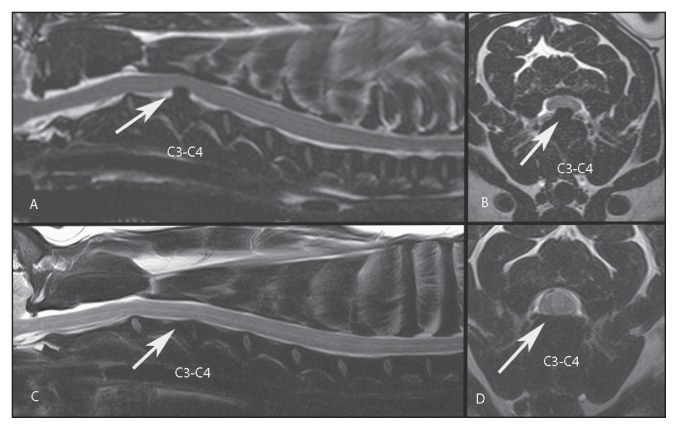
This report describes, for the first time in small animal literature, the spontaneous resorption of herniated Hansen type I intervertebral disc material in the cervical spine of a chondrodystrophic dog over a 4-month period, documented by magnetic resonance imaging. Clinical signs (cervical hyperpathia) responded to conservative treatment during the same period.
Résorption spontanée d’une hernie discale chez un chien détectée par imagerie par résonance magnétique. Cet article décrit, pour la première fois dans la littérature des petits animaux, la résorption spontanée d’une hernie Hansen de type I du matériel du disque intervertébral dans la colonne cervicale d’un chien chondrodystrophique pendant une période de 4 mois et documentée par imagerie par résonance magnétique (IRM). Les signes cliniques (hyperpathie cervicale) ont répondu à un traitement conservateur durant la même période.(Traduit par Isabelle Vallières).
Figures
Similar articles
-
Detection of spinal cord compression in dogs with cervical intervertebral disc disease by magnetic resonance imaging.Vet Rec. 2008 Jul 5;163(1):11-5. doi: 10.1136/vr.163.1.11. Vet Rec. 2008. PMID: 18603629
-
Clinical and imaging findings in dogs with nerve root signature associated with cervical intervertebral disc herniation.J Vet Intern Med. 2024 Mar-Apr;38(2):1111-1119. doi: 10.1111/jvim.16982. Epub 2024 Jan 12. J Vet Intern Med. 2024. PMID: 38216520 Free PMC article.
-
Effectiveness of cervical hemilaminectomy in canine Hansen Type I and Type II disc disease: a retrospective study.J Am Anim Hosp Assoc. 2011 Sep-Oct;47(5):342-50. doi: 10.5326/JAAHA-MS-5604. Epub 2011 Aug 18. J Am Anim Hosp Assoc. 2011. PMID: 21852506
-
Spontaneous regression of lumbar Hansen type 1 disk extrusion detected with magnetic resonance imaging in a dog.J Am Vet Med Assoc. 2014 Mar 15;244(6):715-8. doi: 10.2460/javma.244.6.715. J Am Vet Med Assoc. 2014. PMID: 24568114 Review.
-
Spontaneous regression of herniated cervical disc.Spine J. 2003 Mar-Apr;3(2):171-3. doi: 10.1016/s1529-9430(02)00556-9. Spine J. 2003. PMID: 14589234 Review.
Cited by
-
Case Report: Dural infiltration of intervertebral disc material mimicking spinal cord meningioma in a dog.Front Vet Sci. 2025 Jul 1;12:1621529. doi: 10.3389/fvets.2025.1621529. eCollection 2025. Front Vet Sci. 2025. PMID: 40666731 Free PMC article.
References
-
- Brisson BA. Intervertebral disc disease in dogs. Vet Clin North Am Small Anim Pract. 2010;40:829–858. - PubMed
-
- Lorenz MD, Coates JR, Kent R. Handbook of Veterinary Neurology. 5th ed. St. Louis, Missouri: Saunders; 2011. pp. 164–188.
-
- Cherrone KL, Dewey CW, Coates JR, Bergman RL. A retrospective comparison of cervical intervertebral disk disease in nonchondrodystrophic large dogs versus small dogs. J Am Anim Hosp Assoc. 2004;40:316–320. - PubMed
-
- Sharp NJH, Wheeler SJ. Small Animal Spinal Disorders: Diagnosis and Surgery. 2nd ed. Philadelphia, Pennsylvania: Mosby; 2005. Thoracolumbar disc disease; p. 125.
-
- Jeffery ND, Levine JM, Olby NJ, Stein VM. Intervertebral disk degeneration in dogs: Consequences, diagnosis, treatment, and future directions. J Vet Intern Med. 2013;27:1318–1333. - PubMed
MeSH terms
LinkOut - more resources
Full Text Sources
Medical