Light Microscopy and Polarized Microscopy: A Dermatological Tool to Diagnose Gray Hair Syndromes
- PMID: 28761265
- PMCID: PMC5514796
- DOI: 10.4103/ijt.ijt_21_16
Light Microscopy and Polarized Microscopy: A Dermatological Tool to Diagnose Gray Hair Syndromes
Abstract
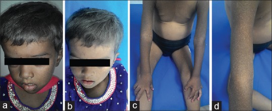
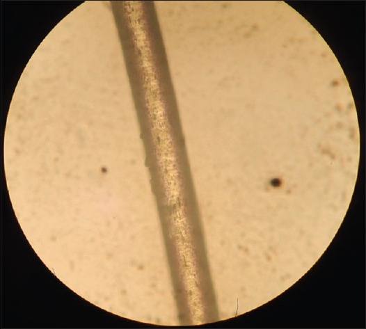
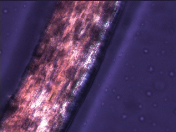
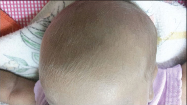
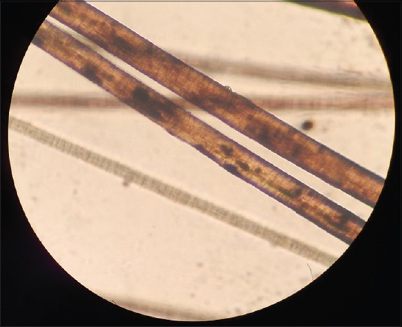
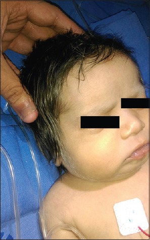
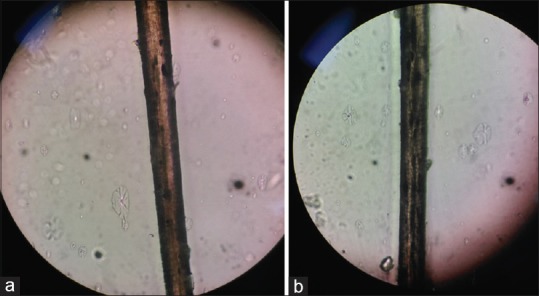
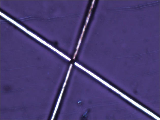
Gray hair syndromes are rare syndromes which have an autosomal recessive inheritance and are characterized by pigmentary dilution of skin and hair, defects in immunological function, and nervous system defects. They comprise three disorders namely Chediak-Higashi syndrome (CHS), Griscelli syndrome (GPS), and Elejalde syndrome. Clinically, it is difficult to distinguish these disorders as their clinical features may overlap. Hence, to make a correct diagnosis and differentiate between CHS and GPS light microscopic examination of skin and hair shafts as well as peripheral blood smear evaluations should be done. In cases where the diagnosis is not possible chromosomal analysis for specific mutations can be done. In resource-poor settings where chromosomal analysis is not possible, and light microscopy findings are inconclusive, polarized microscopy can serve as a useful tool to distinguish between CHS and GPS. We report three cases with gray hair syndromes where the diagnosis on light microscopy and polarized microscopy of hair shaft correlated with the bone marrow examination findings and chromosomal analysis, thus emphasizing the importance of a noninvasive, cost-effective, and time-saving alternative in the diagnosis of these syndromes.
Keywords: Chediak–Higashi syndrome; Griscelli syndrome; gray hair syndromes; polarized microscopy.
Conflict of interest statement
There are no conflicts of interest.
Figures
References
-
- Mancini AJ, Chan LS, Paller AS. Partial albinism with immunodeficiency: Griscelli syndrome: Report of a case and review of the literature. J Am Acad Dermatol. 1998;38(2 Pt 2):295–300. - PubMed
-
- Valente NY, Machado MC, Boggio P, Alves AC, Bergonse FN, Casella E, et al. Polarized light microscopy of hair shafts aids in the differential diagnosis of Chédiak-Higashi and Griscelli-Prunieras syndromes. Clinics (Sao Paulo) 2006;61:327–32. - PubMed
-
- Al-Khenaizan S. Hyperpigmentation in Chediak-Higashi syndrome. J Am Acad Dermatol. 2003;49(5 Suppl):S244–6. - PubMed
Publication types
LinkOut - more resources
Full Text Sources
Other Literature Sources