Case Reports
doi: 10.4103/njms.NJMS_64_16.
Unilateral condylar hyperplasia - A genetic link? Case reports
Affiliations
- PMID: 28761278
- PMCID: PMC5512411
- DOI: 10.4103/njms.NJMS_64_16
Item in Clipboard
Case Reports
Unilateral condylar hyperplasia - A genetic link? Case reports
Natl J Maxillofac Surg.
2017 Jan-Jun.
Abstract
Unilateral condylar hyperplasia is an uncommon condition with unknown etiology which causes overdevelopment of condyle leading to facial asymmetry, mandibular deviation, malocclusion, and articulation dysfunction. Two Indian families with unilateral condylar hyperplasia are presented where the similar abnormality was also detected in one of their parents. The condylar hyperplasia in these two families indicates that mandibular condylar hyperplasia could be genetic in origin.
Keywords: Condylar hyperplasia; facial asymmetry; mandibular condyle.
Conflict of interest statement
There are no conflicts of interest.
Figures

Extraoral photographs of Case 1

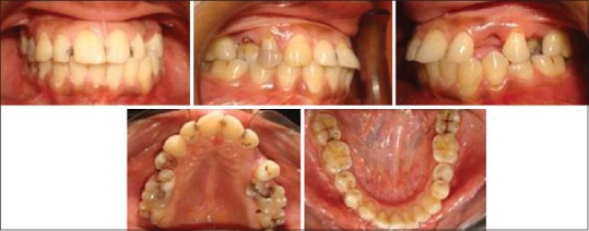
Intraoral photographs of Case 1

Posteroanterior cephalogram of Case 1

Panoramic view of Case 1

Extraoral photographs of mother of Case 1
Intraoral photographs of mother of Case 1

Posteroanterior cephalogram of mother of Case 1

Panoramic view of mother of Case 1

Extraoral photographs of Case 2

Intraoral photographs of Case 2

Panoramic view of Case 2

Posteroanterior cephalogram of Case 2

Extraoral photographs of mother of Case 2

Intraoral photographs of mother of Case 2

Posteroanterior cephalogram of mother of Case 2

Panoramic view of mother of Case 2
Similar articles
-
Ortho-surgical management of condylar hyperplasia: Rare case reports.Natl J Maxillofac Surg. 2014 Jan;5(1):54-9. doi: 10.4103/0975-5950.140180. Natl J Maxillofac Surg. 2014. PMID: 25298720 Free PMC article.
-
Condylar hyperplasia and facial asymmetry: report of five cases.J Maxillofac Oral Surg. 2011 Mar;10(1):50-6. doi: 10.1007/s12663-010-0141-5. Epub 2011 Feb 4. J Maxillofac Oral Surg. 2011. PMID: 22379321 Free PMC article.
-
High condylectomy procedure: a valuable resource for surgical management of the mandibular condylar hyperplasia.J Craniofac Surg. 2013 Jul;24(4):1451-3. doi: 10.1097/SCS.0b013e318285d31f. J Craniofac Surg. 2013. PMID: 23851829
-
[Terminology and classification of condylar hyperplasia: Two case reports and review].Kulak Burun Bogaz Ihtis Derg. 2015;25(6):367-74. doi: 10.5606/kbbihtisas.2015.08068. Kulak Burun Bogaz Ihtis Derg. 2015. PMID: 26572183 Review. Turkish.
-
'Adaptable condylectomy' for acquired facial asymmetry and malocclusion caused by temporomandibular joint condylar hyperplasia.Int J Oral Maxillofac Surg. 2023 Nov;52(11):1145-1155. doi: 10.1016/j.ijom.2023.05.001. Epub 2023 May 23. Int J Oral Maxillofac Surg. 2023. PMID: 37230928 Review.
Cited by
-
Proteomic Expression Profile in Human Temporomandibular Joint Dysfunction.Diagnostics (Basel). 2021 Mar 28;11(4):601. doi: 10.3390/diagnostics11040601. Diagnostics (Basel). 2021. PMID: 33800589 Free PMC article.
References
-
- Nevile BW, Damn DD, Allen CM, Boequot JE, editors. Oral and Maxillofacial Pathology. Philadelphia: Saunders; 1995. pp. 15–6.
-
- de Bont LG, Blankestijn J, Panders AK, Vermey A. Unilateral condylar hyperplasia combined with synovial chondromatosis of the temporomandibular joint. Report of a case. J Oral Maxillofac Surg. 1985;13:32–6. - PubMed
-
- Gray RJ, Sloan P, Quayle AA, Carter DH. Histopathological and scintigraphic features of condylar hyperplasia. J Oral Maxillofac Surg. 1990;19:65–71. - PubMed
-
- Perriman A, Uthman A. Unilateral condylar hyperplasia. Br J Oral Surg. 1971;8:273–80. - PubMed
-
- Obwegeser HL, Makek MS. Hemimandibular hyperplasia – Hemimandibular elongation. J Maxillofac Surg. 1986;14:183–208. - PubMed
Publication types
LinkOut - more resources
Full Text Sources
Other Literature Sources