Suppressive Effects of Mesenchymal Stem Cells in Adipose Tissue on Allergic Contact Dermatitis
- PMID: 28761285
- PMCID: PMC5500702
- DOI: 10.5021/ad.2017.29.4.391
Suppressive Effects of Mesenchymal Stem Cells in Adipose Tissue on Allergic Contact Dermatitis
Abstract
Background: Allergic contact dermatitis (ACD), which is accelerated by interferon (IFN)-γ and suppressed by interleukin (IL)-10 as regulators, is generally self-limited after removal of the contact allergen. Adipose tissue-derived multipotent mesenchymal stem cells (ASCs) potentially exert immunomodulatory effects. Considering that subcutaneous adipose tissue is located close to the site of ACD and includes mesenchymal stem cells (MSCs), the MSCs in adipose tissue could contribute to the self-limiting course of ACD.
Objective: The aims of the present study were to elucidate the effects of MSCs in adipose tissue on ACD and to examine any cytokine-mediated mechanisms involved.
Methods: Ear thickness in a C57BL/6 mouse model of ACD using contact hypersensitivity (CHS) elicited by 2,4,6-trinitro-1-chlorobenzene was evaluated as a marker of inflammation level. Five and nine mice were injected with ASCs and phosphate-buffered saline (PBS), respectively. After ASC or PBS injection, real-time reverse transcription-polymerase chain reaction and enzyme-linked immunosorbent assay were performed.
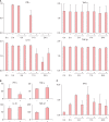
Results: Histology showed that CHS was self-limited and ear thickness was suppressed by ASCs in a dose-dependent manner. IFN-γ expression in the elicited skin site and regional lymph nodes was significantly lower in ASC-treated mice than in control mice. IL-10 expression did not differ between treated and control mice. The suppressive effects of ASCs on CHS response did not differ between IL-10 knock-out C57BL/6 mice and wild-type mice.
Conclusion: The present findings suggest that MSCs in adipose tissue may contribute to the self-limiting course of ACD through decreased expression of IFN-γ, but not through increased expression of IL-10.
Keywords: Adipose tissue; Allergic contact dermatitis; Contact hypersensitivity; Interferon-γ; Interleukin-10; Mesenchymal stem cell.
Conflict of interest statement
CONFLICTS OF INTEREST: The authors have nothing to disclose.
Figures


Similar articles
-
Indoleamine 2,3-Dioxygenase Is Not a Pivotal Regulator Responsible for Suppressing Allergic Airway Inflammation through Adipose-Derived Stem Cells.PLoS One. 2016 Nov 3;11(11):e0165661. doi: 10.1371/journal.pone.0165661. eCollection 2016. PLoS One. 2016. PMID: 27812173 Free PMC article.
-
Initial recruitment of interferon-gamma-producing CD8+ effector cells, followed by infiltration of CD4+ cells in 2,4,6-trinitro-1-chlorobenzene (TNCB)-induced murine contact hypersensitivity reactions.J Dermatol. 2002 Nov;29(11):699-708. doi: 10.1111/j.1346-8138.2002.tb00206.x. J Dermatol. 2002. PMID: 12484431
-
Contribution of human adipose tissue-derived stem cells and the secretome to the skin allograft survival in mice.J Surg Res. 2014 May 1;188(1):280-9. doi: 10.1016/j.jss.2013.10.063. Epub 2014 Jan 27. J Surg Res. 2014. PMID: 24560349
-
Adipose-derived mesenchymal stem cells modulate the immune response in chronic experimental autoimmune encephalomyelitis model.IUBMB Life. 2016 Feb;68(2):106-15. doi: 10.1002/iub.1469. Epub 2016 Jan 12. IUBMB Life. 2016. PMID: 26757144 Review.
-
Application of proteomics in the elucidation of chemical-mediated allergic contact dermatitis.Toxicol Res (Camb). 2017 Jun 13;6(5):595-610. doi: 10.1039/c7tx00058h. eCollection 2017 Sep 1. Toxicol Res (Camb). 2017. PMID: 30090528 Free PMC article. Review.
Cited by
-
An Update on the Potential of Mesenchymal Stem Cell Therapy for Cutaneous Diseases.Stem Cells Int. 2021 Jan 5;2021:8834590. doi: 10.1155/2021/8834590. eCollection 2021. Stem Cells Int. 2021. PMID: 33505474 Free PMC article. Review.
-
Therapeutic potential of adipose derived stromal cells for major skin inflammatory diseases.Front Med (Lausanne). 2024 Feb 23;11:1298229. doi: 10.3389/fmed.2024.1298229. eCollection 2024. Front Med (Lausanne). 2024. PMID: 38463491 Free PMC article. Review.
-
Mesenchymal stem/stromal cell therapy in atopic dermatitis and chronic urticaria: immunological and clinical viewpoints.Stem Cell Res Ther. 2021 Oct 11;12(1):539. doi: 10.1186/s13287-021-02583-4. Stem Cell Res Ther. 2021. PMID: 34635172 Free PMC article. Review.
-
Human Adipose Tissue-Derived Mesenchymal Stem Cells Attenuate Atopic Dermatitis by Regulating the Expression of MIP-2, miR-122a-SOCS1 Axis, and Th1/Th2 Responses.Front Pharmacol. 2018 Nov 6;9:1175. doi: 10.3389/fphar.2018.01175. eCollection 2018. Front Pharmacol. 2018. PMID: 30459600 Free PMC article.
References
-
- Saint-Mezard P, Rosieres A, Krasteva M, Berard F, Dubois B, Kaiserlian D, et al. Allergic contact dermatitis. Eur J Dermatol. 2004;14:284–295. - PubMed
-
- Gaspari AA, Katz SI. Contact hypersensitivity. Curr Protoc Immunol. 2001;(Chapter 4):Unit 4.2. - PubMed
-
- Tončić RJ, Lipozenčić J, Martinac I, Gregurić S. Immunology of allergic contact dermatitis. Acta Dermatovenerol Croat. 2011;19:51–68. - PubMed
-
- Sebastiani S, Albanesi C, De PO, Puddu P, Cavani A, Girolomoni G. The role of chemokines in allergic contact dermatitis. Arch Dermatol Res. 2002;293:552–559. - PubMed
-
- Cavani A, Albanesi C, Traidl C, Sebastiani S, Girolomoni G. Effector and regulatory T cells in allergic contact dermatitis. Trends Immunol. 2001;22:118–120. - PubMed
LinkOut - more resources
Full Text Sources
Other Literature Sources
Molecular Biology Databases
Miscellaneous

