

Selective dorsal rhizotomy: A multidisciplinary approach to treating spastic diplegia
- PMID: 28761524
- PMCID: PMC5532931
- DOI: 10.4103/1793-5482.175625
Selective dorsal rhizotomy: A multidisciplinary approach to treating spastic diplegia
Abstract
Background: Spasticity is a motor disorder that interferes with mobility and affects the quality of life. Different approaches have been utilized to address patients with spastic diplegia, among which is selective dorsal rhizotomy (SDR). Although SDR has been shown to be efficacious in treating spastic patients, many neurologists and neurosurgeons are not well aware of the procedure, its indications, and expected outcomes due to the limited number of centers performing this procedure.
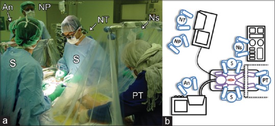
Objectives: The aim of this study is to describe the collaborative multidisciplinary approach between neurosurgeons, neurophysiologists, and physiotherapists in performing SDR. In addition, we delineate three illustrative cases in which SDR was performed in our patients.
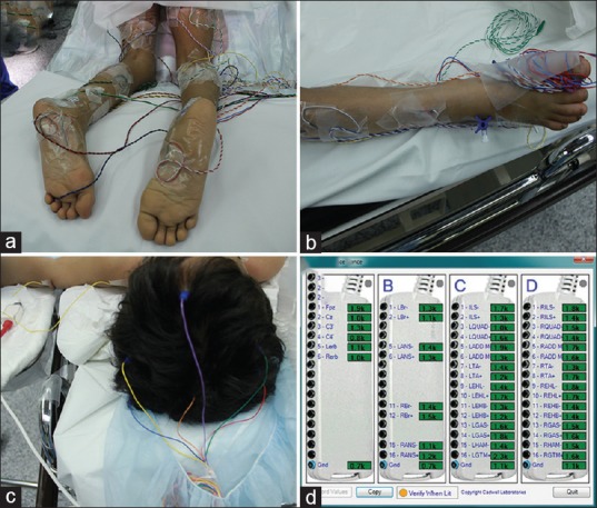
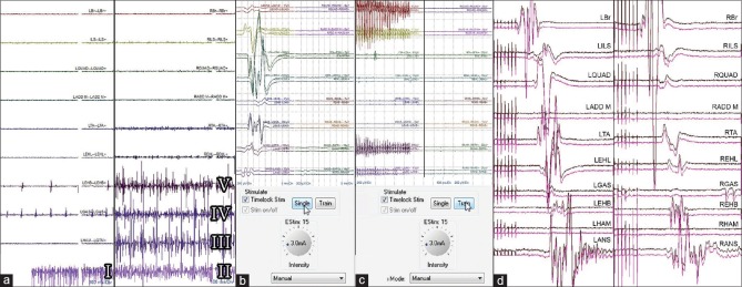
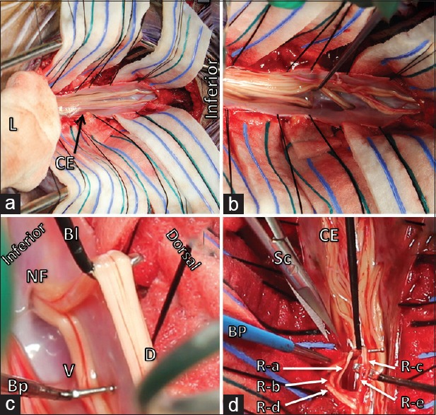
Materials and methods: A retrospective review and analysis of the clinical records of our three patients who underwent SDR was conducted and reported. Patients' outcomes were evaluated and compared to preoperative measurements based on clinical examination of power, tone (Ashworth scale), gait, and range of motion, as well as subjective functional assessment, gross motor function classification system, and gross motor function measure with follow-up at 6, 12, and 24 months postoperatively. A detailed description of our neurosurgical technique in performing SDR in collaboration with neurophysiology and physiotherapy monitoring is provided.
Results: The three patients who underwent SDR using our multidisciplinary approach improved both functionally and objectively after the procedure. No intraoperative or postoperative complications were encountered. All patients were doing well over a long postoperative follow-up period.
Conclusion: A multidisciplinary approach to treating spastic diplegia with SDR can provide good short-term and long-term outcomes in select patients suffering from spastic diplegia.
Keywords: Cerebral palsy; intraoperative monitoring; neurophysiology; physiotherapy; selective dorsal rhizotomy; spastic diplegia; spasticity.
Conflict of interest statement
There are no conflicts of interest.
Figures
References
-
- Prevalence and characteristics of children with cerebral palsy in Europe. Dev Med Child Neurol. 2002;44:633–40. - PubMed
-
- Cahill-Rowley K, Rose J. Etiology of impaired selective motor control: Emerging evidence and its implications for research and treatment in cerebral palsy. Dev Med Child Neurol. 2014;56:522–8. - PubMed
-
- Krigger KW. Cerebral palsy: An overview. Am Fam Physician. 2006;73:91–100. - PubMed
-
- Farmer JP, Sabbagh AJ. Selective dorsal rhizotomies in the treatment of spasticity related to cerebral palsy. Childs Nerv Syst. 2007;23:991–1002. - PubMed
-
- du Plessis AJ, Volpe JJ. Perinatal brain injury in the preterm and term newborn. Curr Opin Neurol. 2002;15:151–7. - PubMed
LinkOut - more resources
Full Text Sources
Other Literature Sources
Miscellaneous