. 2017 Jun 30:11:54-65.
doi: 10.2174/1874104501711010054. eCollection 2017.

Toxicity and Apoptosis Related Effects of Benzimidazo [3,2-α] Quinolinium Salts Upon Human Lymphoma Cells

Affiliations

Toxicity and Apoptosis Related Effects of Benzimidazo [3,2-α] Quinolinium Salts Upon Human Lymphoma Cells

Christian Vélez et al. Open Med Chem J. .

Abstract

Objectives: The present study evaluates novel cationic quinoline derivatives known as benzimidazo[3,2-a]quinolinium salts (BQS) named NBQ-48 and ABQ-48 that have structural similarities to known anti-cancer substances such as ellipticine and berberine.

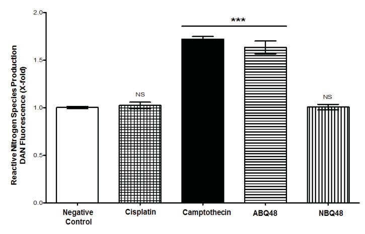
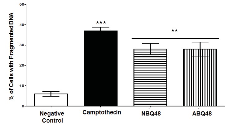
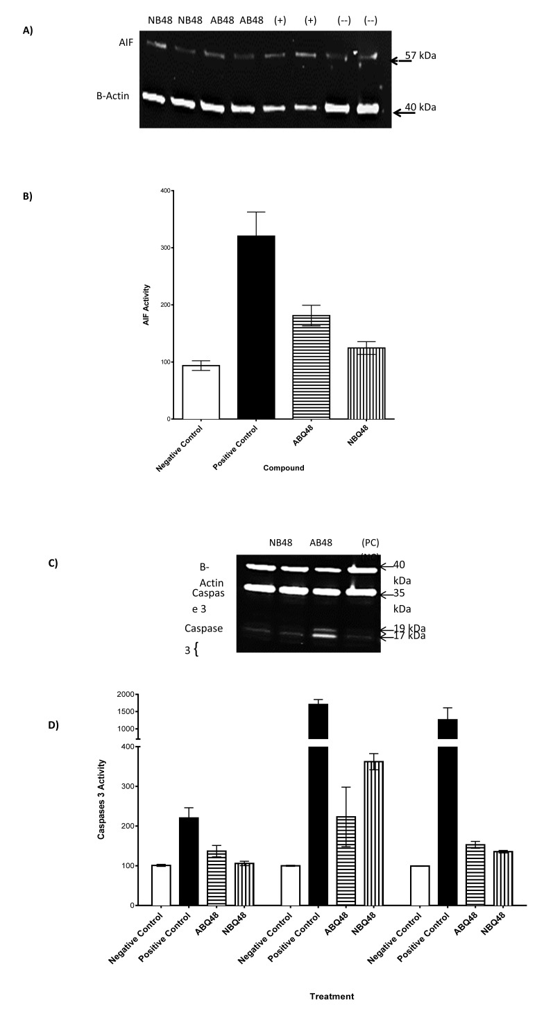
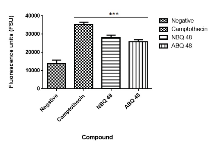
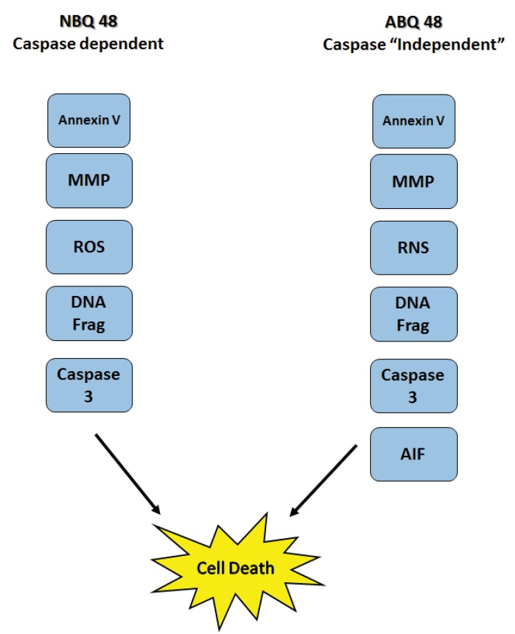
Methods: Toledo human lymphoma (ATCC CRL2631) cells were treated for 24 to 48 hours. Apoptosis related endpoints such as cell cycle arrest, mitochondrial damage, RNS and ROS generation and the activity of several apoptosis related proteins such as caspases and apoptosis inducing factor (AIF) were studied using fluorescence staining and western blot respectively.

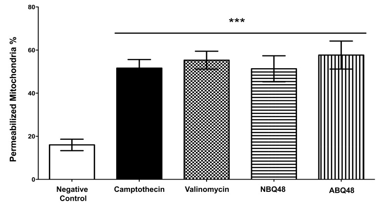
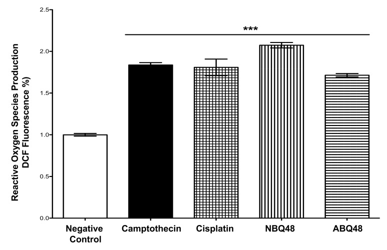
Results: Results indicated a higher toxicity from the amino substituted ABQ-48 versus the NBQ-48 (GI50's of 50uM versus 100uM respectively). Both compounds induced cell death through various apoptosis related endpoints including a decrease in mitochondrial membrane potential with an increase in ROS and activation of the effector caspase 3. Interestingly, AIF release was observed on cells treated with the amino substituted ABQ-48 but not on the nitro substituted NBQ-48 samples suggesting a caspase independent mechanism for ABQ-48.

Conclusions: The results obtained presents the toxic effects of two novel benzimidazo[3,2-a]quinolinium salts in human lymphoma tumor cells. The identified mechanism of action includes multiple apoptosis related effects. Furthermore the data presents a clear variation in caspase dependent or independent mechanism for each compound.

Keywords: Anti cancer; Apoptosis Inducing Factor; Apotosis; BQS; Benzimidazo[3,2-a]quinolinium Salts; Diffuse large B-cell Lymphoma; Unnatural alkaloid.


Figures

Fig. (1a)
Fig. (1a)
General Structure of the two benzimidazo[3,2-a]quinolinium salts studied.
Fig. (1b)
Fig. (1b)
Molecular formula and R1 susbtituents ofstudied compounds. NBQ-48 presents a nitro substituted moiety at the R1 position whereas ABQ-48 has an amino group.
Fig. (2)
Fig. (2)
Mitochondrial membrane permeabilization. Results present permeabilization of cells exposed to NBQ-48 and ABQ-48. A 51.3% of Cells treated with NBQ-48 presented permeabilized membrane versus 57.67% of ABQ-48 treated cells presented mitochondrial membrane permeabilization. Positive controls Camptothecin and Valinomycin presented 51.6% and 55.3% respectively. Clear and significant statistical difference was observed on mitochondrial membrane permeabilization among the tested compounds and the negative control (P<0.05).
Fig. (3)
Fig. (3)
ROS levels were determined after 24h exposure to the negative control (vehicle), NBQ-48 (100 µM), ABQ-48 (50 µM), camptothecin (CP, positive control, 50 µM), and Cisplatin (CS, positive control, 40 µM). Cells exposed to ABQ-48 and NBQ-48 showed significant (p<0.0001) increased ROS levels in comparison to negative control as determined by ANOVA. Statistical analyses performed were a one-way ANOVA, with Tukey post hoc test, where p <0.05 was considered to be significant. P summary; significantly higher in contrast to the negative control (p <0.0001).
Fig. (4)
Fig. (4)
RNS production after 24 h of exposure to vehicle (negative control), ABQ-48 (50 µM), NBQ-48 (100 µM), camptothecin (PC, positive control, 50 µM) and Cisplatin (CS, positive control, 40 µM) was determined. Cells exposed to ABQ-48, and PC exhibited significant induction of reactive nitrogen species (RNS) production on CRL2631 cells when compared to the negative control. Statistical analysis performed was one way ANOVA, with Tukey post hoc test, where p <0.05 was considered to be significant. P summary; significantly different from negative control group (p <0.0001), ns = no significant difference from negative control group (p > 0.05). Results percent (%) was calculated by negative control normalization.
Fig. (5)
Fig. (5)
DNA Fragmentation were determined after 24 h of exposure to negative control (vehicle), NBQ-48 (100 µM), ABQ-48 (50 µM), and camptothecin (CP, positive control, 50 µM). Cells treated with: both NBQ-48 and ABQ-48 showed 28% percent of fragmented DNA and CP presented a 37% cells with fragmented DNA. ANOVA Statistical analisis showed significant (p <0.001) difference in cells exposed to CP, ABQ-48 and NBQ-48 when compared with the negative control. Experiment was performed in replicates and normalized with negative control (expressed in percent %). Statistical analysis performed was a one-way ANOVA, with Tukey post hoc test, where p <0.05 was considered to be significant. (P summary;*** = p <0.0001, ** p<0.001).
Fig. (6)
Fig. (6)
Realease of AIF and activation of caspases after ABQ-48 and NBQ-48 24 hr treatments. (A) Western blot shows lysates of Toledo non-Hodgkin's B cell lymphoma cell line (ATCC CRL2631). A specific band was detected for AIF at approximately 57 kDa (as indicated). (B) At least three independent experiments were performed and data shown are the mean±SD (n=6). ***p < 0.0001 compared to the drugs treated group. (C) Western blot analysis of caspase 3 and 8, results showed presence of caspases 3. (D) Activity of caspase 3 was detected, significant changes were observed in Caspase-3 antibody endogenous levels of full length caspase-3 (35 kDa) and the large fragments of caspase-3 resulting from cleavage (17 and 19 kDa).
Fig. (7)
Fig. (7)
Annexin V assay. Negative sample presented the lowest level of apoptosis induction while the highest level was observed in the positive camptothecin control. Both experimental BQ compounds presented apoptosis levels comparable to the positive control. Statistical analysis performed was a one-way ANOVA, with Tukey post hoc test, where p <0.05 was considered to be significant. (P summary;*** = p <0.0001).
Fig. (8)
Fig. (8)
Summary of detected apoptosis related events on Toledo Lymphoma cells after treatment with ABQ-48 and NBQ-48. Results suggest difference in the mechanisms of action. ABQ-48 induced caspase independent with probable downstream caspase activation while NBQ-48 can induce caspase dependent cell death. Mitochondrial membrane permeabilization (MMP), reactive oxygen species (ROS), reactive nitrogen species (RNS), DNA fragmentation (DNA Frag), Apoptosis inducing factor (AIF).

References

    1. Friedberg Jonathan. Diffuse Large B-Cell Lymphoma. Hematol Oncol. Clin. North Hematol Oncol. Clin North Am. 2008;22(5):941–ix. doi: 10.1016/j.hoc.2008.07.002.. October. - DOI - PMC - PubMed
    1. Flowers C.R, Sinha R, Vose J.M. Improving outcomes for patients with diffuselarge B-cell lymphoma. CA Cancer J. Clin. 2010;60(6):393–408. doi: 10.3322/caac.20087.. Nov-Dec, Epub 2010 Oct 28. - DOI - PubMed
    1. Hurren R., Beheshti Zavareh R., Dalili S., Wood T., Rose D., Chang H., et al. A novel diquinolonium displays preclinical anti-cancer activity and induces caspase-independent cell death. Apoptosis : An Inter. J. on Programmed Cell Death. 2008;13(6):748–755. doi: 10.1007/s10495-008-0209-6. - DOI - PubMed
    1. Cox O., Jackson H., Vargas V.A., Báez A., Colón J.I., González B.C., de León M. Synthesis and Biological Activity of Benzothiazolo- and Benzoxazolo[3,2-a]quinolinium Salts. J. Med. Chem. 1982;25(11):1378–1381. doi: 10.1021/jm00353a020. - DOI - PubMed
    1. Casiano C.A., Báez A. Effects of the Antitumor Drugs 3-Nitrobenzothiazolo[3,2-alpha]quinolinium and Fa- garonine on Nucleic Acid and Protein Synthesis. Biochem. Pharmacol. 1986;35(4):679–685. doi: 10.1016/0006-2952(86)90367-9. - DOI - PubMed

LinkOut - more resources