. 2018 Apr;38(3):679-690.
doi: 10.1007/s10571-017-0527-8. Epub 2017 Jul 31.

Inhibition of Autophagy is Involved in the Protective Effects of Ginsenoside Rb1 on Spinal Cord Injury

Affiliations

Inhibition of Autophagy is Involved in the Protective Effects of Ginsenoside Rb1 on Spinal Cord Injury

Peng Wang et al. Cell Mol Neurobiol. 2018 Apr.

Abstract

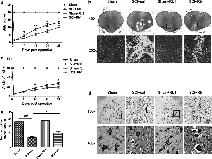
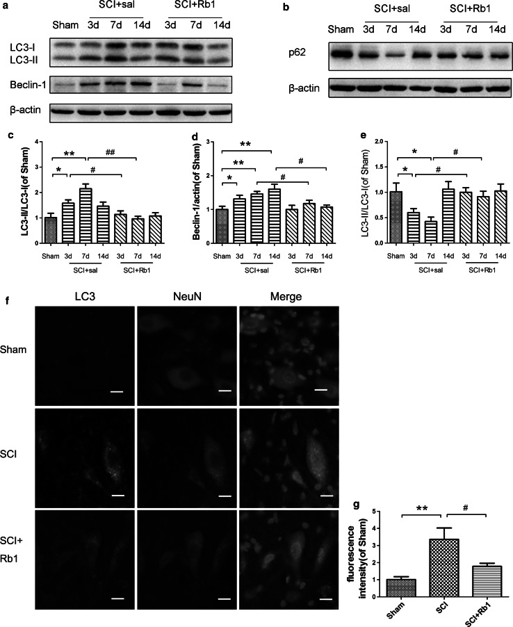
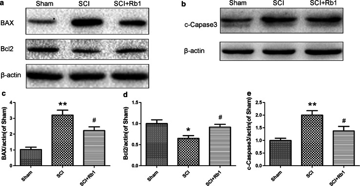
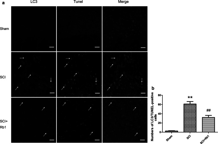
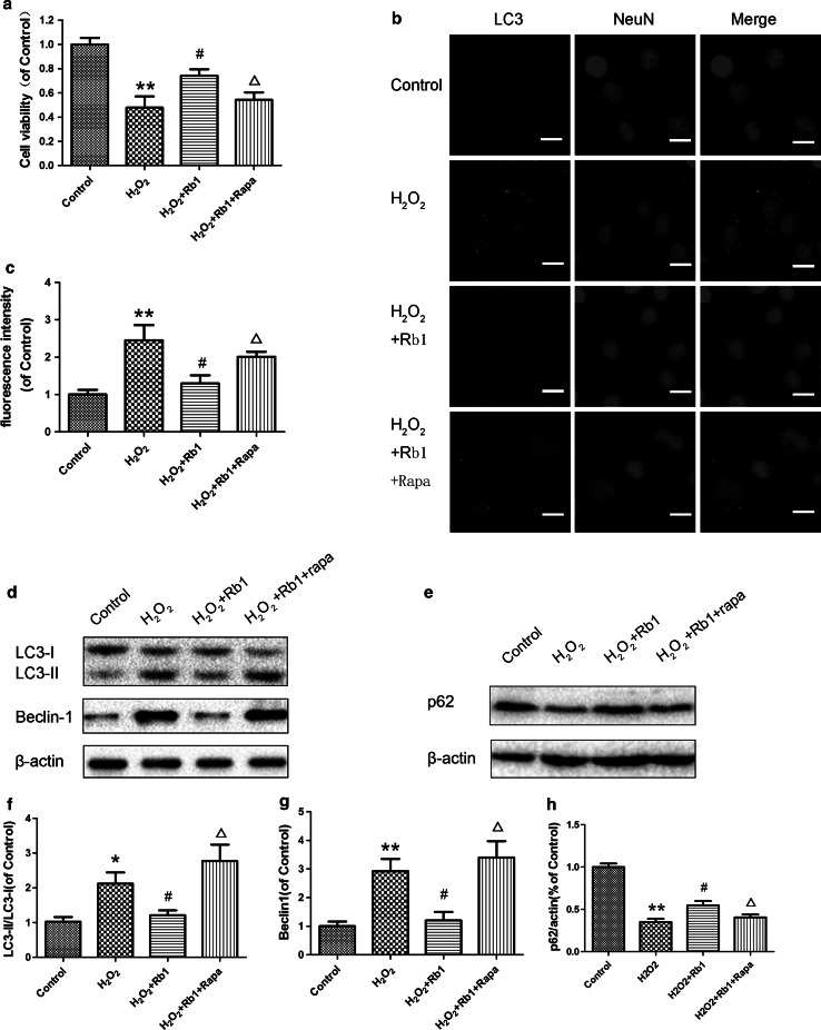
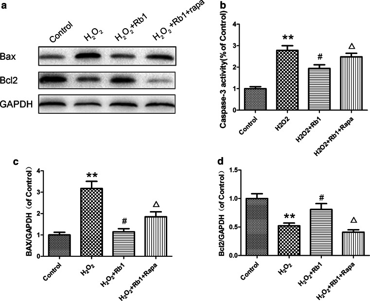
Spinal cord injury (SCI) is a devastating neurological disorder. Autophagy is induced and plays a crucial role in SCI. Ginsenoside Rb1 (Rb1), one of the major active components extracted from Panax Ginseng CA Meyer, has exhibited neuroprotective effects in various neurodegenerative diseases. However, it remains unknown whether autophagy is involved in the neuroprotection of Rb1 on SCI. In this study, we examined the regulation of autophagy following Rb1 treatment and its involvement in the Rb1-induced neuroprotection in SCI and in vitro injury model. Firstly, we found that Rb1 treatment decreased the loss of motor neurons and promoted function recovery in the SCI model. Furthermore, we found that Rb1 treatment inhibited autophagy in neurons, and suppressed neuronal apoptosis and autophagic cell death in the SCI model. Finally, in the in vitro injury model, Rb1 treatment increased the viability of PC12 cells and suppressed apoptosis by inhibiting excessive autophagy, whereas stimulation of autophagy by rapamycin abolished the anti-apoptosis effect of Rb1. Taken together, these findings suggest that the inhibition of autophagy is involved in the neuroprotective effects of Rb1 on SCI.

Keywords: Apoptosis; Autophagy; Ginsenoside Rb1; Spinal cord injury.


Conflict of interest statement

The authors declare that there is no conflict of interest.

Figures

Fig. 1
Fig. 1
Rb1 decreases motor neurons loss and promotes function recovery in the SCI model. a and c the Basso, Beattie and Bresnahan (BBB) scores and angles of inclined plane of rats in sham group, SCI group and SCI + Rb1 group at 0, 1, 7, 14, 21, and 28 days after SCI. b Representative HE staining of the spinal cords in four groups at 28 days after SCI. Magnification is ×40 and ×200, respectively. d Representative Nissl staining of ventral horn of the spinal cords at 28 days after SCI. Magnification is ×200 and ×400, respectively. e Histograms of the number of Nissl positive cells in ventral horn of the spinal cord. Data are mean ± SD, n = 6 per group. ** P < 0.01, * P < 0.05 versus the sham group, # P < 0.05 versus the SCI + saline group
Fig. 2
Fig. 2
Rb1 inhibits autophagy in neurons in the SCI model. At 3, 7, and 14 days after SCI, the expression of LC3-II/I, Beclin-1, and p62 was detected in the spinal cord lesion epicenter of rats treated with Rb1 or saline and sham group. a and b Representative western blot bands of LC3-II/I, Beclin-1, and p62. β-actin was used as the loading control. ce Histograms of the relative expression of LC3-II/I, Beclin-1, and p62. f Representative immunofluorescent staining of LC3 (Green) and NeuN (Red),while nuclei are labeled with DAPI. Scale bars are 10 μm. g Fluorescence intensity of LC3 signal. Data are mean ± SD, n = 6 per group. * P < 0.05, ** P < 0.01 versus the sham group, # P < 0.05, ## P < 0.01 versus the SCI + saline group (Color figure online)
Fig. 3
Fig. 3
Rb1 inhibits apoptosis in the SCI Model. a and b Representative western blot bands of Bax, Bcl-2 and c-Caspase3 in the spinal cord lesion epicenter at 7 days after SCI. β-actin was used as the loading control. ce Histograms of the relative expression of Bax, Bcl-2 and c-Caspase3 versus β-actin. Data are mean ± SD, n = 6 per group. **P < 0.01, *P < 0.05 versus Sham group, # P < 0.05 versus the SCI + saline group
Fig. 4
Fig. 4
Rb1 suppresses autophagic cell death induced by SCI. a Representative double-immunofluorescence analysis of LC3 (green) and TUNEL (red) in ventral horn area at 7 days after SCI. Scale bars are 50 μm. b Counting of LC3 and TUNEL-positive cells among groups. Data are mean ± SD, n = 6 per group. ** P < 0.01, versus Sham group, ## P < 0.05 versus the SCI + saline group (Color figure online)
Fig. 5
Fig. 5
Rb1 decreases H2O2-induced cell death via autophgay inhibition in vitro. PC12 cells were treated as described in the text. a Protective effects of Rb1 on H2O2-induced decrease in PC12 cell viability by CCK8 assay. b and c Immunofluorescent staining of LC3 (Green), while the nuclei are labeled with DAPI (blue). Scale bars are 10 μm. d and e Representative western blot bands of LC3-II/I, Beclin-1, and p62 in PC12 cells. β-actin was used as the loading control. fh Histograms of the relative expression of LC3-II/I, Beclin-1, and p62 versus β-actin. Data are mean ± SD, n = 5 per group. * P < 0.05, ** P < 0.01 versus control group, # P < 0.05 versus the H2O2 group, P < 0.05 versus the H2O2 +Rb1 group (Color figure online)
Fig. 6
Fig. 6
Rb1 decreases H2O2-induced cell death via autophgay inhibition in vitro. a Representative western blot bands of Bax and Bcl-2 in PC12 cells of four groups. β-actin was used as the loading control. cd Histograms of the relative expression of Bax and Bcl-2 versus β-actin. b The effect of Rb1 on caspase-3 activity in H2O2-induced PC12 cells injury. Caspase-3 activity was detected with a commercial kit following the manufacturer’s instruction. Data are mean ± SD, n = 5 per group. ** P < 0.01 versus control group, # P < 0.05 versus the H2O2 group, P < 0.05 versus the H2O2 +Rb1 group

Similar articles

Cited by

References

    1. Adhami F, Liao G, Morozov YM, Schloemer A, Schmithorst VJ, Lorenz JN, Dunn RS, Vorhees CV, Wills-Karp M, Degen JL, Davis RJ, Mizushima N, Rakic P, Dardzinski BJ, Holland SK, Sharp FR, Kuan CY (2006) Cerebral ischemia-hypoxia induces intravascular coagulation and autophagy. Am J Pathol 169(2):566–583. doi:10.2353/ajpath.2006.051066 - PMC - PubMed
    1. Bisicchia E, Latini L, Cavallucci V, Sasso V, Nicolin V, Molinari M, D’Amelio M, Viscomi MT (2016) Autophagy inhibition favors survival of rubrospinal neurons after spinal cord hemisection. Mol Neurobiol. doi:10.1007/s12035-016-0031-z - PubMed
    1. Bursch W, Ellinger A, Kienzl H, Torok L, Pandey S, Sikorska M, Walker R, Hermann RS (1996) Active cell death induced by the anti-estrogens tamoxifen and ICI 164 384 in human mammary carcinoma cells (MCF-7) in culture: the role of autophagy. Carcinogenesis 17(8):1595–1607 - PubMed
    1. Chen HC, Fong TH, Lee AW, Chiu WT (2012) Autophagy is activated in injured neurons and inhibited by methylprednisolone after experimental spinal cord injury. Spine 37(6):470–475. doi:10.1097/BRS.0b013e318221e859 - PubMed
    1. Chen B, Yue R, Yang Y, Zeng H, Chang W, Gao N, Yuan X, Zhang W, Shan L (2015) Protective effects of (E)-2-(1-hydroxyl-4-oxocyclohexyl) ethyl caffeine against hydrogen peroxide-induced injury in PC12 cells. Neurochem Res 40(3):531–541. doi:10.1007/s11064-014-1498-5 - PubMed

LinkOut - more resources