

Sentinel Lymph Node Characterization with a Dual-Targeted Molecular Ultrasound Contrast Agent
- PMID: 28762204
- PMCID: PMC6535621
- DOI: 10.1007/s11307-017-1109-3
Sentinel Lymph Node Characterization with a Dual-Targeted Molecular Ultrasound Contrast Agent
Abstract
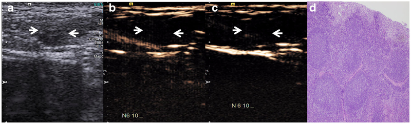
Purpose: The purpose of this study was to assess the performance of molecular ultrasound with dual-targeted microbubbles to detect metastatic disease in the sentinel lymph nodes (SLNs) in swine model of naturally occurring melanoma. The SLN is the first lymph node in the lymphatic chain draining primary tumor, and early detection of metastatic SLN involvement is critical in the appropriate management of melanoma.
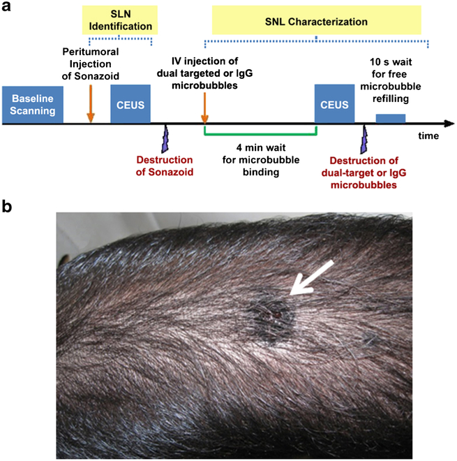
Procedure: Nine Sinclair swine (weight 3-7 kg; Sinclair BioResources, Columbia, MO, USA) with naturally occurring melanoma were examined. Siemens S3000 scanner with a 9L4 probe was used for imaging (Siemens Healthineers, Mountain View, CA). Dual-targeted contrast agent was created using Targestar SA microbubbles (Targeson, San Diego, CA, USA) labeled with ανβ3-integrin and P-selectin antibodies. Targestar SA microbubbles labeled with IgG-labeled were used as control. First, peritumoral injection of Sonazoid contrast agent (GE Healthcare, Oslo, Norway) was performed to detect SLNs. After that, dual-targeted and IGG control Targestar SA microbubbles were injected intravenously with a 30-min interval between injections. Labeled Targestar SA microbubbles were allowed to circulate for 4 min to enable binding. After that, two sets of image clips were acquired several seconds before and after a high-power destruction sequence. The mean intensity difference pre- to post-bubble destruction within the region of interest placed over SLN was calculated as a relative measure of targeted microbubble contrast agent retention. This process was repeated for non-SLNs as controls. All lymph nodes evaluated on imaging were surgically removed and histologically examined for presence of metastatic involvement.
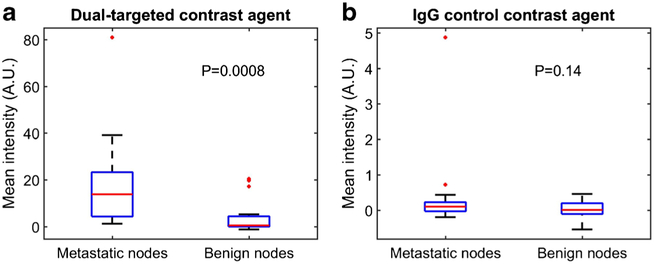
Results: A total of 43 lymph nodes (25 SLNs and 18 non-SLNs) were included in the analysis with 18 SLNs demonstrating metastatic involvement greater than 5 % on histology. All non-SLNs were benign. The mean intensity (± SD) of the dual-targeted microbubbles for metastatic SLNs was significantly higher than that of benign LNs (18.05 ± 19.11 vs. 3.30 ± 6.65 AU; p = 0.0008), while IgG-labeled control microbubbles demonstrated no difference in retained contrast intensity between metastatic and benign lymph nodes (0.39 ± 1.14 vs. 0.03 ± 0.24 AU; p = 0.14).
Conclusions: The results indicate that dual-targeted microbubbles labeled with P-selectin and ανβ3-integrin antibodies may aid in detecting metastatic involvement in SLNs of melanoma.
Keywords: Angiogenesis; Characterization; Contrast imaging; Inflammation; Melanoma; Metastasis; Sentinel lymph nodes; Targeted microbubbles; Ultrasound.
Conflict of interest statement
Conflict of Interest
Dr. Forsberg is on the Speaker’s Bureau of GE Healthcare, and Dr. Eisenbrey receives funding, equipment, and drug support from GE Healthcare. The other authors declare that they have no relevant conflict of interest.
Figures
References
-
- American Cancer Society (2017) Melanoma skin cancer. http://www.cancer.org/cancer/skincancer-melanoma. Accessed 6 April 2017
-
- Morton DL, Thompson JF, Cochran AJ et al. (2006) Sentinel-node biopsy or nodal observation in melanoma. N Engl J Med 355:1307–1317 - PubMed
Publication types
MeSH terms
Substances
Grants and funding
LinkOut - more resources
Full Text Sources
Other Literature Sources
Research Materials
