. 2017 Aug 1;18(8):1666.
doi: 10.3390/ijms18081666.

Toll-Like Receptor-4 Inhibitor TAK-242 Attenuates Motor Dysfunction and Spinal Cord Pathology in an Amyotrophic Lateral Sclerosis Mouse Model

Affiliations

Toll-Like Receptor-4 Inhibitor TAK-242 Attenuates Motor Dysfunction and Spinal Cord Pathology in an Amyotrophic Lateral Sclerosis Mouse Model

Avi Fellner et al. Int J Mol Sci. .

Abstract

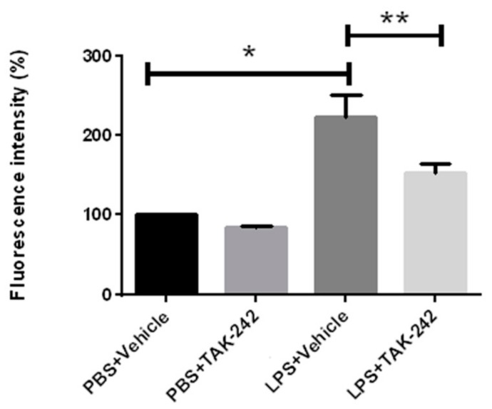
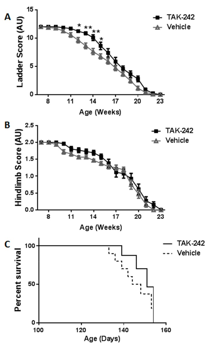
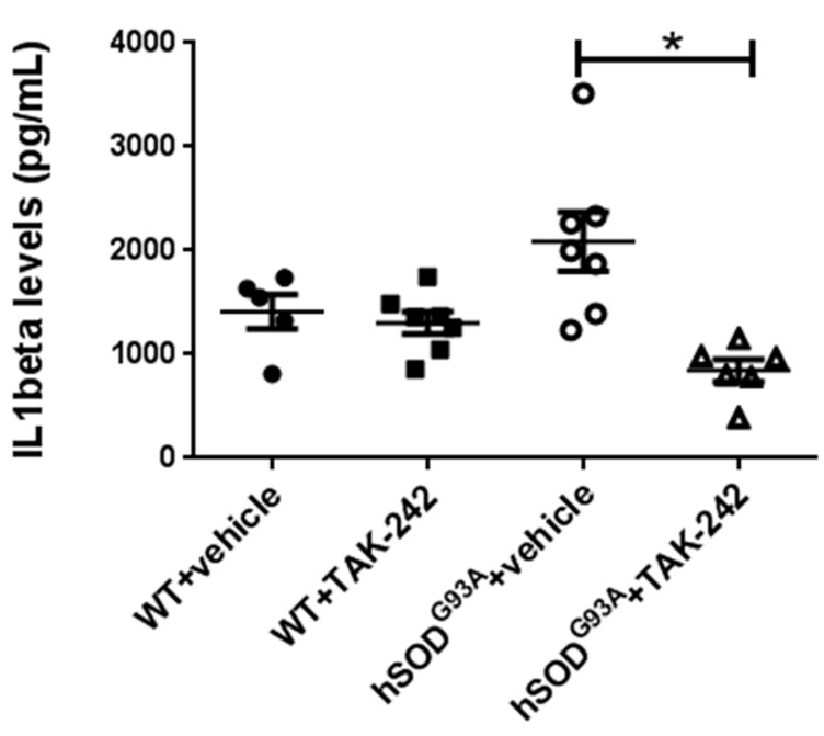
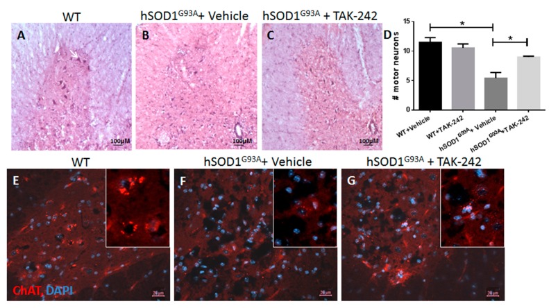
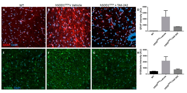
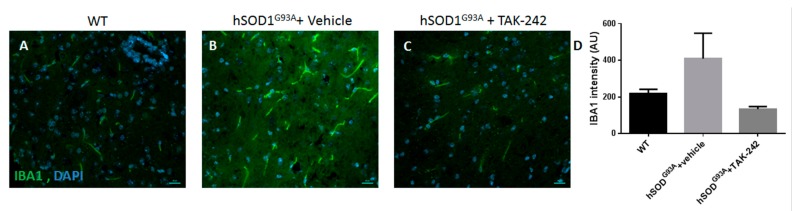
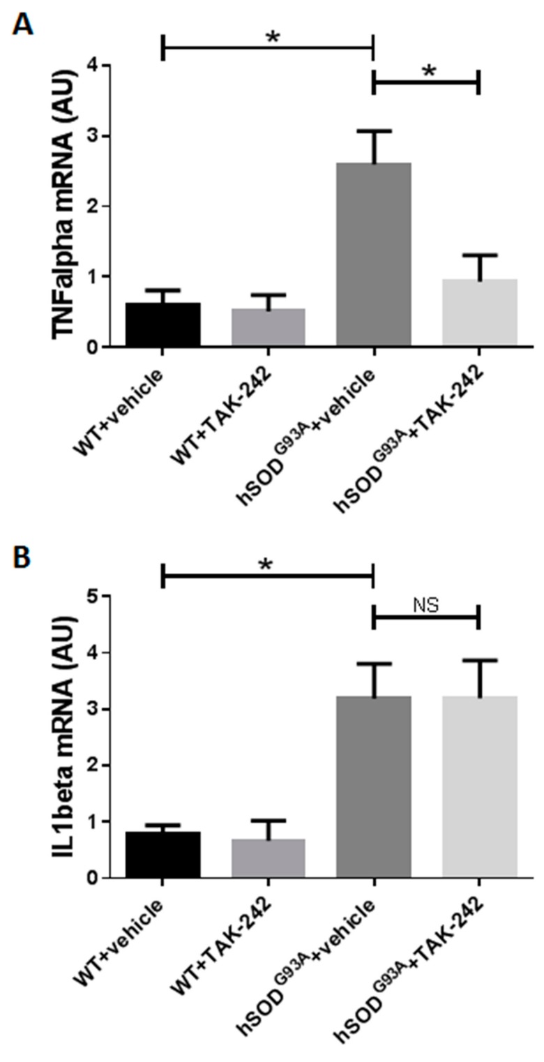
Neuroinflammation contributes to amyotrophic lateral sclerosis (ALS) progression. TLR4, a transmembrane protein that plays a central role in activation of the innate immune system, has been shown to induce microglial activation in ALS models. TLR4 is up-regulated in the spinal cords of hSOD1G93A mice. We aimed to examine the effects of specific TLR4 inhibition on disease progression and survival in the hSOD1G93A mouse model of ALS. Immunologic effect of TLR4 inhibition in vitro was measured by the effect of TAK-242 treatment on LPS-induced splenocytes proliferation. hSOD1G93A transgenic mice were treated with TAK-242, a selective TLR4 inhibitor, or vehicle. Survival, body weight, and motor behavior were monitored. To evaluate in vivo immunologic modifications associated with TAK-242 treatment, we measured serum IL-1β in the plasma, as well as IL-1β and TNF-α mRNAs in the spinal cord in wild-type mice and in TAK-242-treated and vehicle-treated early symptomatic hSOD1G93A mice. Immunohistochemical analysis of motor neurons, astrocytes, and microglial reactivity in the spinal cords were performed on symptomatic (100 days old) TAK-242-treated and vehicle-treated hSOD1G93A mice. In vitro, splenocytes taken from 100 days old hSOD1G93A mice showed significantly increased proliferation when exposed to LPS (p = 0.0002), a phenomenon that was reduced by TAK-242 (p = 0.0179). TAK-242 treatment did not attenuate body weight loss or significantly affect survival. However, TAK-242-treated hSOD1G93A mice showed temporary clinical delay in disease progression evident in the ladder test and hindlimb reflex measurements. Plasma IL-1β levels were significantly reduced in TAK-242-treated compared to vehicle-treated hSOD1G93A mice (p = 0.0023). TAK-242 treatment reduced spinal cord astrogliosis and microglial activation and significantly attenuated spinal cord motor neuron loss at early disease stage (p = 0.0259). Compared to wild-type animals, both IL-1β and TNF-α mRNAs were significantly upregulated in the spinal cords of hSOD1G93A mice. Spinal cord analysis in TAK-242-treated hSOD1G93A mice revealed significant attenuation of TNF-α mRNA (p = 0.0431), but no change in IL-1β mRNA. TLR4 inhibition delayed disease progression, attenuated spinal cord astroglial and microglial reaction, and reduced spinal motor neuron loss in the ALS hSOD1G93A mouse model. However, this effect did not result in increased survival. To our knowledge, this is the first report on TAK-242 treatment in a neurodegenerative disease model. Further studies are warranted to assess TLR4 as a therapeutic target in ALS.

Keywords: ALS; SOD1 mice; TAK-242; TLR4; animal model; innate immune system; neuroinflammation.


Conflict of interest statement

The authors declare no conflict of interest.

Figures

Figure 1
Figure 1
LPS-induced splenocyte proliferation in vitro was significantly reduced by TAK-242 treatment. Splenocytes obtained from 100 days old hSODG93A mice were treated with TAK-242 or vehicle for three days. Proliferation was measured from the Alamar blue assay and quantified by fluorescence intensity. PBS: phosphate-buffered saline; LPS: lipopolysaccharide. Data are expressed as mean ± SEM, * p < 0.01, ** p < 0.05.
Figure 2
Figure 2
Behavioral motor performance analysis showed delay in disease progression in TAK-242- treated hSODG93A mice. (A) The ladder test. A score of 12 represents completely healthy mice and 0 correlates with disease end stage. Ladder testing showed statistically significant differences between hSODG93A mice treated with TAK-242 or vehicle. Data are expressed as mean ± SEM, * p < 0.05, ** p < 0.01; (B) A similar trend in disease progression was found in hind limb reflex score testing. Data are expressed as mean ± SEM; (C) Survival analysis by Kaplan–Meier curves of TAK-242 and vehicle-treated hSODG93A mice. All wild-type littermates survived and kept having a normal maximal score in all behavioral tests during the whole experiment period (not shown in the graphs).
Figure 3
Figure 3
Prolonged TAK-242 treatment attenuates serum IL1-β levels of early symptomatic hSODG93A mice. IL1-β levels were measured by ELISA testing in sera obtained at day 100 of life, after 50 days of treatment with TAK-242 or vehicle. Analysis showed an increase in IL1-β levels in vehicle-treated hSODG93A mice compared to WT mice, which was significantly attenuated by TAK-242 treatment. WT: wild type. * p < 0.01.
Figure 4
Figure 4
Preservation of spinal motor neurons in TAK-242-treated hSODG93A mice. Lumbar spinal cords sections (9 sections from each animal; 3 animals in each group) from early symptomatic hSODG93A mice (100 days old) were stained by H&E staining (AC); Immunohistochemistry staining for the motor neuron marker ChAT is shown in (EG); Motor neuron number was significantly reduced in hSODG93A mice treated with vehicle (B,F) compared to WT mice (A,E); Motor neuron loss was significantly reduced in hSODG93A mice treated with TAK-242 (C,G); Motor neurons quantification is shown in (D) and represents average number of motor neurons per one ventral horn in each group. An example of a motor neuron is marked by the arrow in A. WT: wild type; H&E: hematoxylin and eosin; ChAT: choline acetyltransferase; DAPI: 4,6-diamidino-2-phenylindole. Scale bars are 100 µm for (AC) and 20 µm for (EG). Data are expressed as mean ± SEM, * p < 0.05.
Figure 5
Figure 5
Prolonged TAK-242 treatment attenuates astroglial activation in hSODG93A mice spinal cords. Immunohistochemistry staining for the astrocyte markers GFAP (red; AC) and S100β (green; EG) at day 100 of life showed increased spinal astrogliosis in vehicle-treated hSODG93A mice (B,F) compared to WT littermates (A,E), that was prominently attenuated in TAK-242 treated hSODG93A mice (C (p = 0.0462); G (p = 0.0481)). Quantification of staining intensity is shown graphically in panels D (GFAP) and H (S100β). WT: wild type; GFAP: glial fibrillary acidic protein; DAPI: 4,6-diamidino-2-phenylindole. Scale bars for all panels = 20 µm.
Figure 6
Figure 6
Prolonged TAK-242 treatment attenuates microglial activation in hSODG93A mice spinal cords. Immunohistochemistry staining for the activated microglia marker IBA1 at day 100 of life showed increased spinal microglial activation in vehicle-treated hSODG93A mice (B) as compared to WT littermates (A). Microglial activation was prominently attenuated by prolonged TAK-242 treatment of hSODG93A mice (C; p = 0.0544). Quantification of staining intensity is shown graphically in panel (D). WT: wild type; IBA1: ionized calcium-binding adapter molecule-1; DAPI: 4,6-diamidino-2-phenylindole. Scale bars for all panels = 20 µm.
Figure 7
Figure 7
Prolonged TAK-242 treatment attenuated TNF-α expression but not IL1-β expression in the spinal cords of early symptomatic hSODG93A mice. TNF-α (A) and IL1-β (B) mRNA levels in the spinal cords of TAK-242-treated and vehicle-treated 100-days old WT and early symptomatic hSODG93A mice were quantified by real-time PCR as described in the methods section. WT: wild type; NS: non-significant. Data are expressed as mean ± SEM, * p < 0.05.

Similar articles

Cited by

References

    1. Ropper A.H., Samuels M.A., Klein J.P., editors. Adams and Victor’s Principles of Neurology. 10th ed. McGraw-Hill; New York, NY, USA: 2014. Degenerative diseases of the nervous system; pp. 1109–1116.
    1. Lee J.Y., Lee J.D., Phipps S., Noakes P.G., Woodruff T.M. Absence of toll-like receptor 4 (TLR4) extends survival in the hSOD1G93A mouse model for amyotrophic lateral sclerosis. J. Neuroinflamm. 2015 doi: 10.1186/s12974-015-0310-z. - DOI - PMC - PubMed
    1. Kiernan M.C., Vucic S., Cheah B.C., Turner M.R., Eisen A., Hardiman O., Zoing M.C. Amyotrophic lateral sclerosis. Lancet. 2013;377:942–955. doi: 10.1016/S0140-6736(10)61156-7. - DOI - PubMed
    1. Arjoud-Driss S., Siddique T. Sporadic and hereditary amyotrophic lateral sclerosis (ALS) Biochim. Biophys. Acta. 2015;1852:679–684. doi: 10.1016/j.bbadis.2014.08.010. - DOI - PubMed
    1. Gonzales H., Elgueta D., Montoya A., Pachero R. Neuroimmune regulation of microglial activity involved in neuroinflammation and neurodegenerative diseases. J. Neuroimmunol. 2014;274:1–13. doi: 10.1016/j.jneuroim.2014.07.012. - DOI - PubMed

MeSH terms

Substances