. 2017 Aug 1;12(8):e0181868.
doi: 10.1371/journal.pone.0181868. eCollection 2017.

RORγt and RORα signature genes in human Th17 cells

Affiliations

RORγt and RORα signature genes in human Th17 cells

Glenda Castro et al. PLoS One. .

Abstract

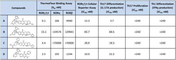
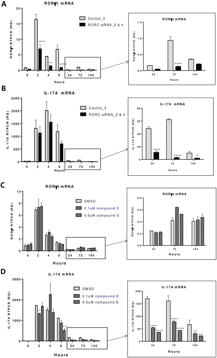
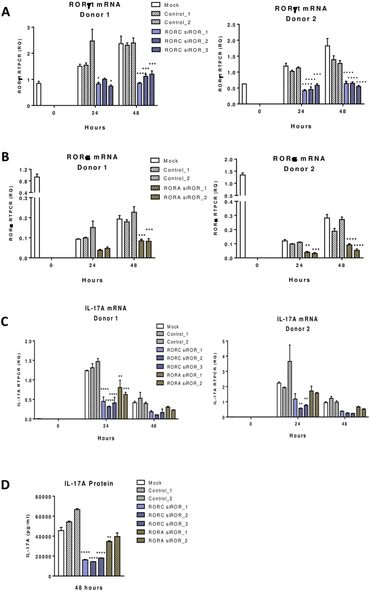
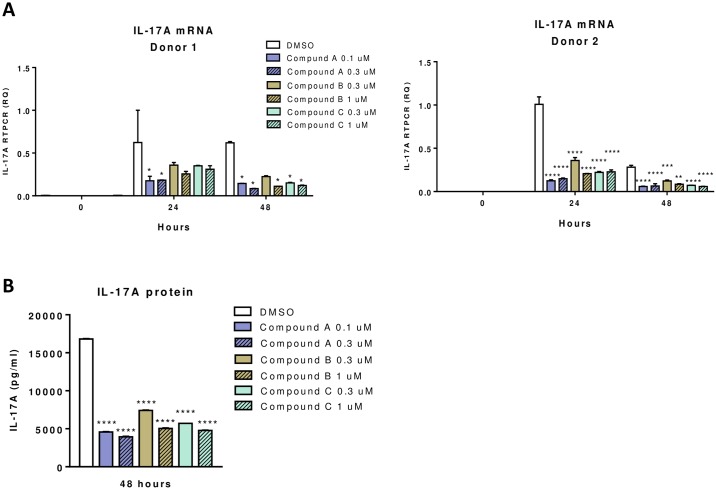
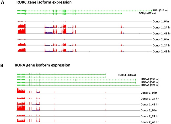
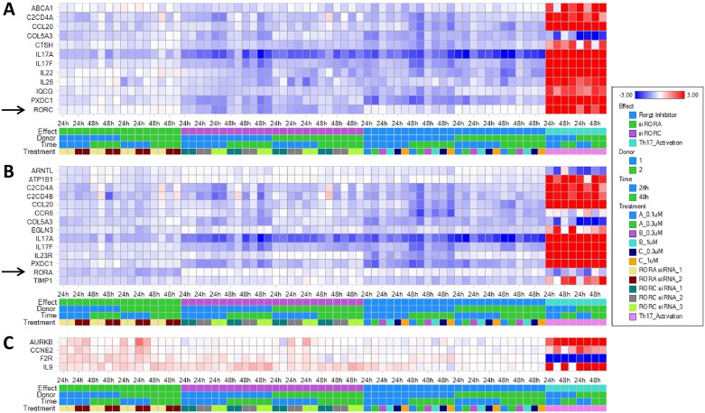
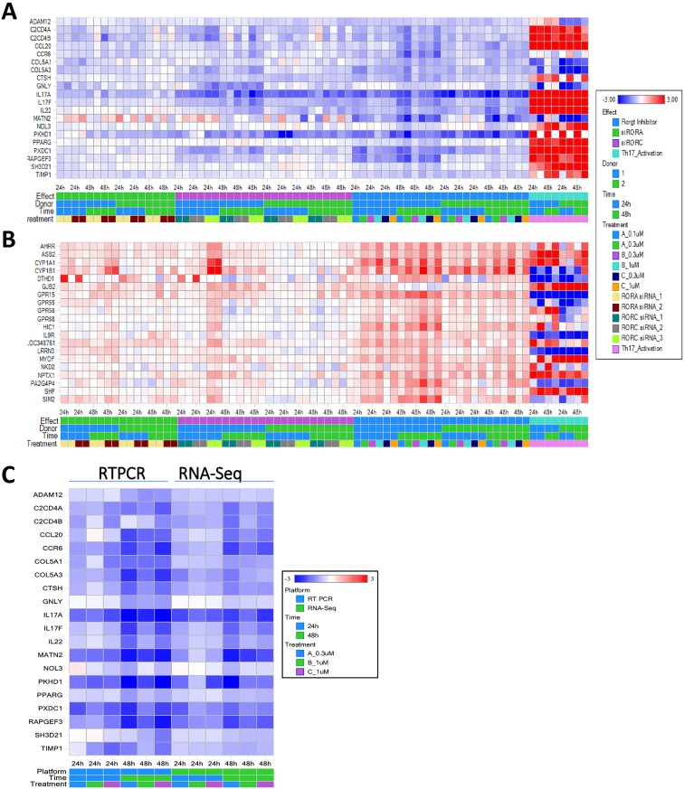
RORγt and RORα are transcription factors of the RAR-related orphan nuclear receptor (ROR) family. They are expressed in Th17 cells and have been suggested to play a role in Th17 differentiation. Although RORγt signature genes have been characterized in mouse Th17 cells, detailed information on its transcriptional control in human Th17 cells is limited and even less is known about RORα signature genes which have not been reported in either human or mouse T cells. In this study, global gene expression of human CD4 T cells activated under Th17 skewing conditions was profiled by RNA sequencing. RORγt and RORα signature genes were identified in these Th17 cells treated with specific siRNAs to knock down RORγt or RORα expression. We have generated selective small molecule RORγt modulators and they were also utilized as pharmacological tools in RORγt signature gene identification. Our results showed that RORγt controlled the expression of a very selective number of genes in Th17 cells and most of them were regulated by RORα as well albeit a weaker influence. Key Th17 genes including IL-17A, IL-17F, IL-23R, CCL20 and CCR6 were shown to be regulated by both RORγt and RORα. Our results demonstrated an overlapping role of RORγt and RORα in human Th17 cell differentiation through regulation of a defined common set of Th17 genes. RORγt as a drug target for treatment of Th17 mediated autoimmune diseases such as psoriasis has been demonstrated recently in clinical trials. Our results suggest that RORα could be involved in same disease mechanisms and gene signatures identified in this report could be valuable biomarkers for tracking the pharmacodynamic effects of compounds that modulate RORγt or RORα activities in patients.


Conflict of interest statement

Competing Interests: All authors are employees of Janssen R&D. The RORgt inverse agonists were generated by Janssen R&D and will require material transfer agreements prior to sharing but this does not alter our adherence to PLOS ONE policies on sharing data and materials.

Figures

Fig 1
Fig 1. RORγt inverse agonists used in gene signature studies.
Compound structures and their potencies in binding assay, cellular reporter assay, and T cell differentiation assay are shown. These compounds were selective in binding to RORγ/γt over RORα and RORβ, effective in blocking RORγt driven luciferase expression, inhibiting Th17 differentiation but not proliferation, and had no effect on Th1 differentiation.
Fig 2
Fig 2. IL-17A transcript in human CD4 T cells reduced by RORγt siRNA or inverse agonist at later but not early stage of Th17 differentiation.
Human CD4 T cells were activated under Th17 differentiation conditions as described in Material and Methods. RORγt siRNAs (RORC siRNA_2 & 4) and control scramble siRNA (control_3) were transfected into CD4 T cells prior to Th17 differentiation and samples were collected at different time points and the expression of RORγt (A) and IL-17A (B) transcripts was measured by RT-PCR as described in Materials and Methods. RORγt compound D at 0.1 and 0.5 μM and DMSO vehicle control were tested in a similar manner in Th17 differentiation. RORγt (C) and IL-17A (D) transcripts were measured by RT-PCR at different time points. Statistical significance of RORC siRNA or compound treated samples in reducing RORγt or IL-17A mRNA at different time points was analyzed with 2-way ANOVA and P values were indicated as *P<0.05, **P<0.01, ***P<0.001 and ****P<0.0001.
Fig 3
Fig 3. RORγt or RORα siRNA effect on IL-17A transcript in human CD4 T cells during Th17 differentiation.
Human CD4 T cells purified from 2 donors were activated under Th17 differentiation condition as described in Material and Methods. RORγt and RORα siRNAs, as well as control siRNAs were transfected into CD4 T cells prior to Th17 differentiation and samples were collected at different time points as indicated. Expression of RORγt (A), RORα (B) and IL-17A (C) transcripts was measured by RT-PCR. IL-17A cytokine released in culture supernatants (D) was measured by ELISA. Statistical significance of the difference between RORC or RORA siRNA treated samples compared to mock samples at different time points was analyzed with 2-way ANOVA and P values were presented as *P<0.05, **P<0.01, ***P<0.001, and ****P<0.0001.
Fig 4
Fig 4. RORγt inverse agonist effect on IL-17A transcript in human CD4 T cells during Th17 differentiation.
Human CD4 T cells purified from 2 donors were activated under Th17 differentiation condition as described in Material and Methods. CD4 T cells were treated with three RORγt inverse agonists at two different concentrations as well as DMSO vehicle control. Expression of IL-17A mRNA in different treated samples was measured by RT-PCR at time points as indicated (A). IL-17A cytokine released in culture supernatants (B) was measured by ELISA. Statistical significance of the difference between compound treated to DMSO samples at different time points was analyzed with 2-way ANOVA and P values were indicated as *P<0.05, **P<0.01, ***P<0.001 and ****P<0.0001.
Fig 5
Fig 5. Expression of RORγt and RORα4 in human CD4 T cells during Th17 differentiation.
Expression of RORγt isoform from the RORC gene (A) and RORα4 isoform from the RORA gene (B) was determined from detection of isoform specific exon sequences in RNA-Seq data analysis. Exons utilized by different isoforms of the RORC and RORA genes are shown in green, and the frequency of exon sequences in RNA-Seq data are shown in red in the histograms. DMSO treated human CD4 T cell samples from 2 donors at different time points of activation under Th17 differentiation conditions were used for isoform expression analysis.
Fig 6
Fig 6. Global gene expression profile with RNA-Seq identified genes differentially regulated by RORγt or RORα siRNA in human CD4 T cells during Th17 differentiation.
RNA sequencing profile was performed on RNA samples extracted from human CD4 T cells treated with RORγt or RORα siRNA, or RORγt inverse agonists as described in S1 Table. Genes down-regulated by RORγt (A) and RORα (B) siRNAs, or up-regulated by RORγt or RORα siRNAs (C) were identified in analysis of RNA-Seq data using the criteria described in Materials and Methods. Each column represents log2 ratios of selected genes in one of the 72 comparisons, including comparison of untreated Th17 cells with RORγt or RORα siRNA transfected cells or RORγt inverse agonist treated cells at two time points and T cells from 2 independent donors.
Fig 7
Fig 7. Global gene expression profile with RNA-Seq identified genes down- or up-regulated by RORγt inverse agonists in human CD4 T cells during Th17 differentiation.
RNA sequencing profile was performed on RNA samples extracted from human CD4 T cells treated with 3 different RORγt inverse agonists at 2 concentrations as described in S1 Table. Genes down-regulated (A) or up-regulated (B) by RORγt inverse agonists were identified in analysis of RNA-Seq data using the criteria described in Materials and Methods. Each column represents log2 ratios of selected genes in one of the 72 comparisons, including comparison of untreated Th17 cells with RORγt or RORα siRNA transfected cells or RORγt inverse agonist treated cells at two time points and T cells from 2 independent donors. Genes down-regulated by RORγt inverse agonists in Th17 cells identified from RNA-Seq analysis were confirmed by RTPCR of donor 1 samples as described in Materials and Methods (C). Heat map of log2 ratios of gene expression in response to treatment of RORγt inverse agonists determined by RNA-Seq and RTPCR were compared.

References

    1. Jetten AM (2009) Retinoid-related orphan receptors (RORs): critical roles in development, immunity, circadian rhythm, and cellular metabolism. Nucl Recept Signal 7: e003 doi: 10.1621/nrs.07003 - DOI - PMC - PubMed
    1. Giguere V, Tini M, Flock G, Ong E, Evans RM, et al. (1994) Isoform-specific amino-terminal domains dictate DNA-binding properties of ROR alpha, a novel family of orphan hormone nuclear receptors. Genes Dev 8: 538–553. - PubMed
    1. Matysiak-Scholze U, Nehls M (1997) The structural integrity of ROR alpha isoforms is mutated in staggerer mice: cerebellar coexpression of ROR alpha1 and ROR alpha4. Genomics 43: 78–84. - PubMed
    1. Carlberg C, Hooft van Huijsduijnen R, Staple JK, DeLamarter JF, Becker-Andre M (1994) RZRs, a new family of retinoid-related orphan receptors that function as both monomers and homodimers. Mol Endocrinol 8: 757–770. - PubMed
    1. Villey I, de Chasseval R, de Villartay JP (1999) RORgammaT, a thymus-specific isoform of the orphan nuclear receptor RORgamma / TOR, is up-regulated by signaling through the pre-T cell receptor and binds to the TEA promoter. Eur J Immunol 29: 4072–4080. - PubMed

MeSH terms

Substances