. 2017 Aug 1;12(8):e0181783.
doi: 10.1371/journal.pone.0181783. eCollection 2017.

Synthesis of novel coumarin nucleus-based DPA drug-like molecular entity: In vitro DNA/Cu(II) binding, DNA cleavage and pro-oxidant mechanism for anticancer action

Affiliations

Synthesis of novel coumarin nucleus-based DPA drug-like molecular entity: In vitro DNA/Cu(II) binding, DNA cleavage and pro-oxidant mechanism for anticancer action

Saman Khan et al. PLoS One. .

Abstract

Despite substantial research on cancer therapeutics, systemic toxicity and drug-resistance limits the clinical application of many drugs like cisplatin. Therefore, new chemotherapeutic strategies against different malignancies are needed. Targeted cancer therapy is a new paradigm for cancer therapeutics which targets pathways or chemical entities specific to cancer cells than normal ones. Unlike normal cells, cancer cells contain elevated copper which plays an integral role in angiogenesis. Copper is an important metal ion associated with chromatin DNA, particularly with guanine. Thus, targeting copper via copper-specific chelators in cancer cells can serve as an effective anticancer strategy. New pharmacophore di(2-picolyl)amine (DPA)-3(bromoacetyl) coumarin (ligand-L) was synthesized and characterized by IR, ESI-MS, 1H- and 13C-NMR. Binding ability of ligand-L to DNA/Cu(II) was evaluated using a plethora of biophysical techniques which revealed ligand-L-DNA and ligand-L-Cu(II) interaction. Competitive displacement assay and docking confirmed non-intercalative binding mode of ligand-L with ctDNA. Cyclic voltammetry confirmed ligand-L causes quasi reversible Cu(II)/Cu(I) conversion. Further, acute toxicity studies revealed no toxic effects of ligand-L on mice. To evaluate the chemotherapeutic potential and anticancer mechanism of ligand-L, DNA damage via pBR322 cleavage assay and reactive oxygen species (ROS) generation were studied. Results demonstrate that ligand-L causes DNA cleavage involving ROS generation in the presence of Cu(II). In conclusion, ligand-L causes redox cycling of Cu(II) to generate ROS which leads to oxidative DNA damage and pro-oxidant cancer cell death. These findings will establish ligand-L as a lead molecule to synthesize new molecules with better copper chelating and pro-oxidant properties against different malignancies.


Conflict of interest statement

Competing Interests: The authors have declared that no competing interests exist.

Figures

Fig 1
Fig 1. Scheme 1: Synthetic route for the synthesis of ligand-L.
Fig 2
Fig 2. Absorption spectra of ligand-L (5 μM) in the absence and presence of ct-DNA (0–35 μM) and Cu(II) (0–35 μM).
(A) Increasing concentration of ct-DNA showed hyperchromic shift suggesting ct-DNA-Ligand-L interaction. (B) Increasing concentration of Cu(II) showed hyperchromic shift suggesting Cu(II)- Ligand-L interaction.
Fig 3
Fig 3
Fluorescence emission spectra of ligand-L (5 μM) in the absence and presence of ct-DNA (0–40 μM) (A) and Cu(II) (0–40 μM) (B). Increasing concentrations of DNA and Cu(II) lead to quenching in fluorescence intensity of ligand-L.
Fig 4
Fig 4
Stern-Volmer plots for interaction of ligand-L with ct-DNA (A) and Cu(II) (B).
Fig 5
Fig 5. Competitive displacement assays.
(A) Fluorescence titration of EtBr-DNA complex with ligand-L. EtBr-DNA complex was excited at 471 nm and emission spectra were recorded from 520–700 nm. No change in fluorescence intensity was recorded on addition of increasing concentration of ligand-L. (B) Fluorescence titration of Hoechst-DNA complex with ligand-L. Hoechst-DNA complex was excited at 343 nm and emission spectra were recorded from 400–600 nm. Fluorescence intensity decreases on addition of increasing concentration of ligand-L.
Fig 6
Fig 6. ITC curve (upper panel) and the binding isotherm (lower panel) for ligand-L interaction with ct-DNA at 25°C.
Fig 7
Fig 7. Effect of ligand-L on CD spectra of ctDNA.
CD spectra of ctDNA (50 μM) with varying concentrations of ligand-L (0–100 μM).
Fig 8
Fig 8. KI quenching experiment.
Stern-Volmer plot for fluorescence quenching of ligand-L (30 μM) by KI in the absence and presence of ctDNA (30 μM). Difference in Ksv value (quenching constant) was used to investigate the binding mode of ligand-L with ctDNA. R is the correlation coefficient.
Fig 9
Fig 9. Molecular docked structure of ligand-L complexed with B-DNA.
(A) Surface view interaction of ligand-L with B-DNA. (B) Hydrogen bonding interactions (5) of ligand-L with B-DNA (PDB ID: 1BNA).
Fig 10
Fig 10. Cyclic voltammogram of ligand-L (25 μM) in absence and presence of Cu(II) (25 μM).
Fig 11
Fig 11. Erythrocyte lysis test.
In vitro toxicity measured on treatment with vehicle control (2% DMSO) and increasing concentrations of ligand-L (25–100 μM). Values expressed as mean ± SEM of three independent experiments.
Fig 12
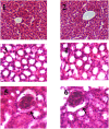
Fig 12. Histological analysis of ligand-L treated groups (H&E staining).
Histological sections of liver (first row) and kidney (second and third row). Untreated (control group) liver (Panel 1) and kidney (Panels 3 and 5) of mice. Liver (Panel 2) and kidney (Panels 4 and 6) from ligand-L (250 mg/kg body weight) treated mice. No significant differences in structures were observed of liver and kidney of control and treated groups. Panels 3 and 4 represent tubular region of control and treated kidney respectively, whereas panels 5 and 6 represent glomerulus region of control and treated kidney respectively.
Fig 13
Fig 13. NBT reduction assay.
(A) Estimation of superoxide anion generation by increasing concentrations of ligand-L (25–100 μM). (B) Superoxide anion generation by ligand-L in the presence of Cu(II) ions and effect of copper chelator (neocuproine) and superoxide dismutase (ROS scavenger) on ROS generation by ligand-L-Cu(II) system. (1) Ligand-L (25 μM) (2) Cu(II) (25 μM) (3) Ligand-L (25 μM) + Cu(II) (25 μM) (4) Ligand-L (25 μM) + Cu(II) (25 μM) + Neocuproine (50 μM) (5) Ligand-L (25 μM) + Cu(II) (25 μM) + SOD (20 μg/ml). *P < 0.05 with respect to ligand-L (25 μM) set and #P < 0.05 with respect to ligand-L (25 μM) + Cu(II) (25 μM) set.
Fig 14
Fig 14
(A) Estimation of hydroxyl radical generation by increasing concentrations of ligand-L (25–100 μM). (B) Hydroxyl radical generation by ligand-L in the presence of Cu(II) ions and effect of copper chelator (neocuproine) and thiourea (ROS scavenger) on ROS generation by ligand-L-Cu(II) system. (1) Control-No treatment (2) Ligand-L (25 μM) (3) Cu(II) (25 μM) (4) Ligand-L (25 μM) + Cu(II) (25 μM) (5) Ligand-L (25 μM) + Cu(II) (25 μM) + Neocuproine (50 μM) (6) Ligand-L (25 μM) + Cu(II) (25 μM) + thiourea (0.2 mM). *P < 0.05 with respect to control and #P < 0.05 with respect to Ligand-L (25 μM) + Cu(II) (25 μM) set.
Fig 15
Fig 15. Cu(II)-Ligand-L system induced plasmid DNA damage.
(A) Treatment of plasmid pBR322 DNA with ligand-L alone (25 μM) (Lane 1) and Cu(II)-Ligand-L system i.e. Ligand-L (25 μM) + Cu(II) (25 μM) (Lane 2), Ligand-L (25 μM) + Cu(II) (50 μM) (Lane 3), Ligand-L (25 μM) + Cu(II) (75 μM) (Lane 4), Ligand-L (25 μM) + Cu(II) (100 μM) (Lane 5). (B) Effect of copper chelator (neocuproine) and ROS scavengers (thiourea, catalase and SOD) on plasmid pBR322 DNA damage induced by ligand-L-Cu(II) system. (Lane1 1): Ligand-L (25 μM) + Cu(II) (25 μM). (Lane 2): Ligand-L (25 μM) + Cu(II) (25 μM) + Neocuproine (50 μM). (Lane 3): Ligand-L (25 μM) + Cu(II) (25 μM) + thiourea (0.2 mM). (Lane 4): Ligand-L (25 μM) + Cu(II) (25 μM) + catalase (20 μg/ml). (Lane 5): Ligand-L (25 μM) + Cu(II) (25 μM) + SOD (20 μg/ml). Lane ‘C’ depicts the ‘Control’ untreated plasmid DNA.

Similar articles

Cited by

References

    1. Hurley LH. DNA and its associated processes as targets for cancer therapy. Nat Rev Cancer. 2002; 2:188–200. doi: 10.1038/nrc749 - DOI - PubMed
    1. Burger RM. Cleavage of nucleic acids by bleomycin. Chem Rev. 1998; 98:1153–1170. - PubMed
    1. Boerner LJ, Zaleski JM. Metal complex-DNA interactions: from transcription inhibition to photoactivated cleavage. Curr Opin Chem Biol. 2005; 9:135–144. doi: 10.1016/j.cbpa.2005.02.010 - DOI - PubMed
    1. Zhang CX, Lippard SJ. New metal complexes as potential therapeutics. Curr Opin Chem Biol. 2003; 7:481–489. - PubMed
    1. Boulikas T, Vougiouka M. Cisplatin and platinum drugs at the molecular level. (Review). Oncol Rep. 2003; 10:1663–1682. - PubMed

MeSH terms

LinkOut - more resources