. 2017 Aug 1;12(8):e0181801.
doi: 10.1371/journal.pone.0181801. eCollection 2017.

Infectious bronchitis corona virus establishes productive infection in avian macrophages interfering with selected antimicrobial functions

Affiliations

Infectious bronchitis corona virus establishes productive infection in avian macrophages interfering with selected antimicrobial functions

Aruna Amarasinghe et al. PLoS One. .

Abstract

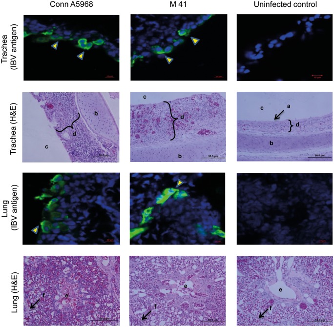
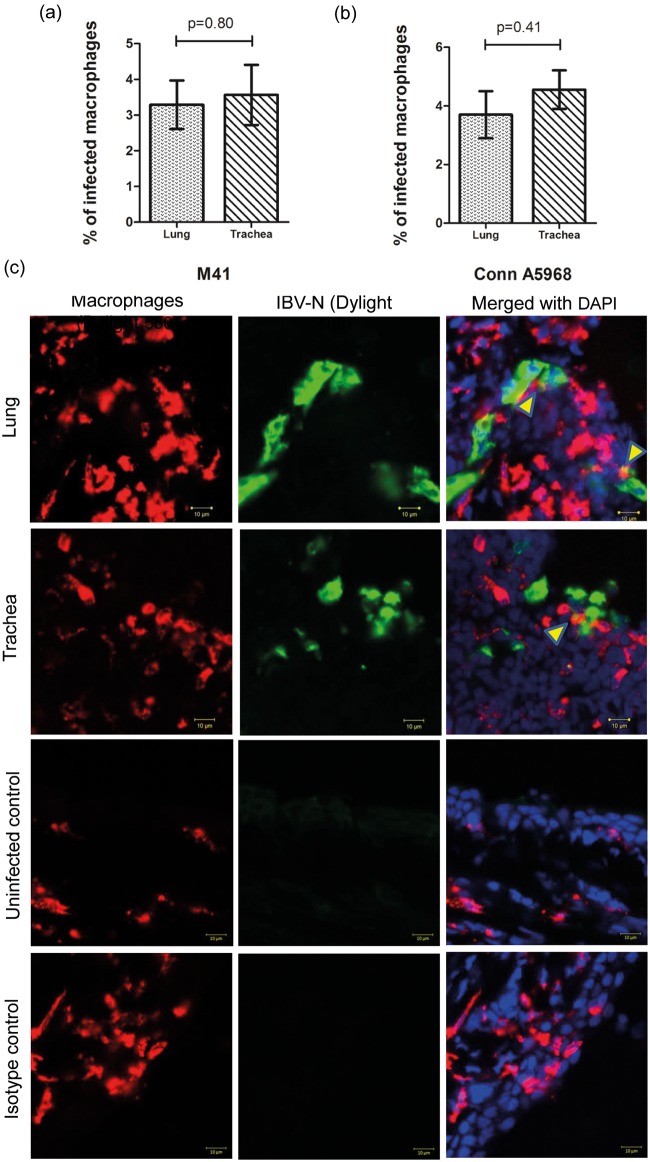
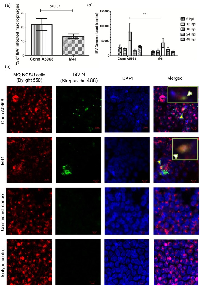
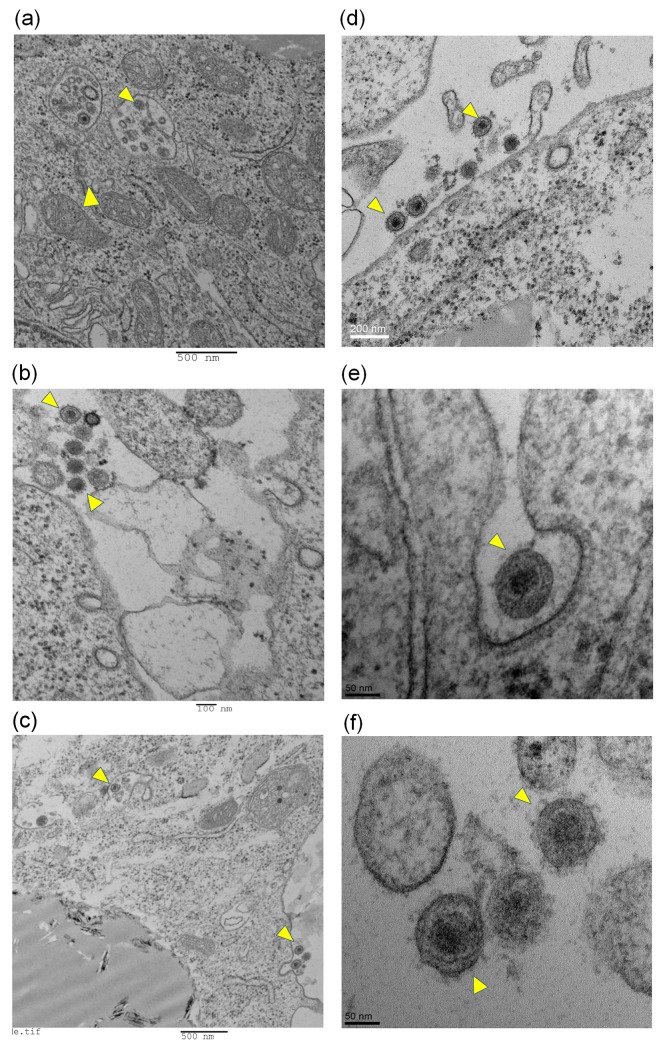
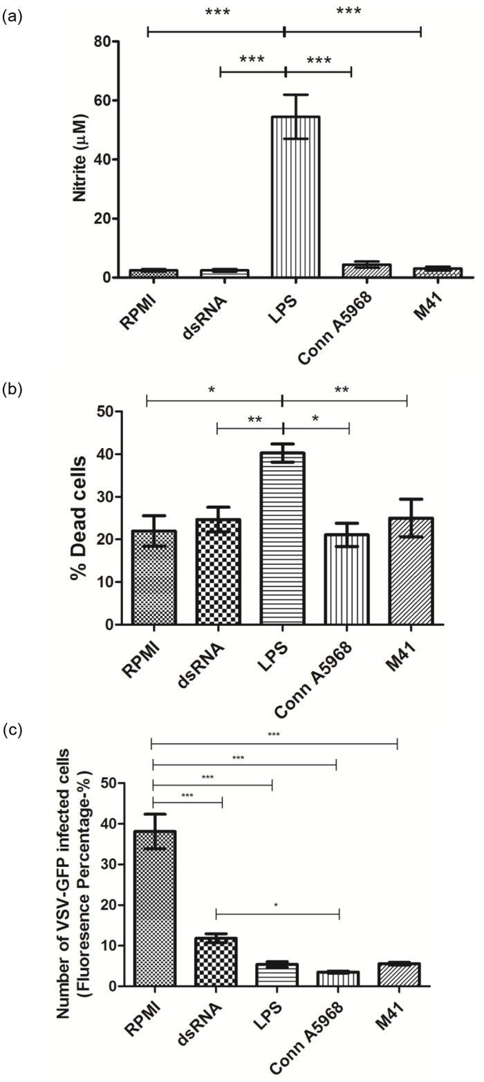
Infectious bronchitis virus (IBV) causes respiratory disease leading to loss of egg and meat production in chickens. Although it is known that macrophage numbers are elevated in the respiratory tract of IBV infected chickens, the role played by macrophages in IBV infection, particularly as a target cell for viral replication, is unknown. In this study, first, we investigated the ability of IBV to establish productive replication in macrophages in lungs and trachea in vivo and in macrophage cell cultures in vitro using two pathogenic IBV strains. Using a double immunofluorescent technique, we observed that both IBV Massachusetts-type 41 (M41) and Connecticut A5968 (Conn A5968) strains replicate in avian macrophages at a low level in vivo. This in vivo observation was substantiated by demonstrating IBV antigens in macrophages following in vitro IBV infection. Further, IBV productive infection in macrophages was confirmed by demonstrating corona viral particles in macrophages and IBV ribonucleic acid (RNA) in culture supernatants. Evaluation of the functions of macrophages following infection of macrophages with IBV M41 and Conn A5968 strains revealed that the production of antimicrobial molecule, nitric oxide (NO) is inhibited. It was also noted that replication of IBV M41 and Conn A5968 strains in macrophages does not interfere with the induction of type 1 IFN activity by macrophages. In conclusion, both M41 and Con A5968 IBV strains infect macrophages in vivo and in vitro resulting productive replications. During the replication of IBV in macrophages, their ability to produce NO can be affected without affecting the ability to induce type 1 IFN activity. Further studies are warranted to uncover the significance of macrophage infection of IBV in the pathogenesis of IBV infection in chickens.


Conflict of interest statement

Competing Interests: The authors have declared that no competing interests exist.

Figures

Fig 1
Fig 1. IBV M41 and Conn A5968 infections were evident by both IBV antigen demonstration and histological changes of the trachea and lungs.
Six days old chickens were infected intra-tracheally with IBV M41 (n = 5) and Conn A5968 (n = 5) strains (2.75×10⁴ EID50 per bird) with uninfected controls (n = 4) and lung and tracheal tissues were collected in OCT and 10% formal saline at 4 dpi. Five μm frozen sections were immunostained for demonstrating IBV antigen and paraffin embedded sections were H&E stained. IBV N antigen in the tracheal and lung sections were stained using DyLight® 550 fluorescent (arrow heads). In the IBV infected trachea, complete loss of cilia (a) and thickening of the mucosa with inflammatory cell infiltration can be seen compared to the control trachea. In the IBV infected lungs, inflammation leading to reduced air exchange areas and reduced lumen of parabronchi are visible. Also, the difference of mucosal thickening in the infected and uninfected trachea is shown (d). a = cilia, b = tracheal cartilage, c = tracheal lumen, d = mucosal layer, e = parabronchial lumen and f = interparabronchial septum.
Fig 2
Fig 2. IBV M41 and Conn A5968 infections increase macrophage number in trachea and lungs.
Six days old SPF chickens were infected intra-tracheally as described in the materials and methods. At 4 dpi, the trachea and lungs were sampled and preserved in OCT for the quantification of macrophage numbers. The 5μm thick frozen sections were stained for macrophages using mouse anti-chicken macrophage primary antibody followed by the secondary anti-mouse antibody conjugated with Dylight 550®. An epifluorescence microscope was used for the quantification of macrophage in immunostained sections. Statistical analysis for identifying group differences was done based on ANOVA test setting the P value <0.05. Trachea and lung macrophage quantitative data, as well as representative immunofluorescent images from each treatment group are given.
Fig 3
Fig 3. Quantification of IBV M41 and Conn A5968 antigens in macrophages in chickens.
Six days old chickens were infected intra-tracheally as indicated in the Fig 1 legend and trachea and lungs were sampled at 4 dpi for double immunostaining of macrophages and IBV antigens. The sections were stained for both macrophages and IBV antigen as has been described in the materials and methods and a confocal microscope was used to obtain representative images. Macrophages were visualized with Dylight 550® (red) and IBV antigens were visualized with Dylight 488® (green). Statistical analysis for identifying group differences was done based on Student’s t test. Nuclei were stained with DAPI (blue). IBV infected macrophages are shown with arrow heads in the merged picture. Both isotype and uninfected controls are shown.
Fig 4
Fig 4. IBV M41 and Conn A5968 strains replicate in macrophages in vitro and establish productive infection.
1.5x 10⁶ of MQ-NCSU cells were cultured on plastic coverslips in 6-well plates for 24 hours before infected with 0.1 MOI of IBV Conn A5968 and M41 strains. After 1 hour of adsorption period, inoculum was drained and cells were washed 2 times with HBSS and incubated at 40°C for another 24 hours. At the end of 24 hours, cell membranes of macrophages were stained with horseradish wheat agglutinin conjugated with Alexa Flour 594. Then the cells were fixed using 4% paraformaldehyde, stained for IBV N antigen and visualized using Dylight 488® conjugated secondary antibody and nuclear staining was done with mounting media with DAPI at the time of mounting coverslips on the slides. (a) Percentage of infected macrophages (MQ-NCSU cells) is shown for each IBV strain. (b) Representative confocal microscopy images showing M41 and Conn A5968 IBV antigens in macrophages. (c) The MQ-NCSU cells were cultured on 6-well plates for 24 hours before infected with 0.1 MOI of IBV Conn A5968 or M41 strains. After 1 hour of adsorption period, inoculum was drained and cells were washed 2 times with HBSS to preclude subsequent quantification of the residual IBV inoculum and continued to incubate at 40°C before collecting cell culture supernatant at 6, 12, 18, 24 and 48 hpi for IBV viral RNA concentration determination using a qPCR assay. All the treatments were done in triplicate and qPCR reactions were carried out in triplicate for each sample as well. For each time point, average number of IBV copies per 200 ng of starting RNA of both IBV Conn A5968 and M41 strains are shown with SEM. ANOVA test was performed to identify group differences and the differences were considered significant at P<0.05. The results represent pooled data of three independent experiments.
Fig 5
Fig 5. Macrophage infections of IBV M41 and Conn A5968 strains produce coronavirus-like particles within macrophages in vitro.
MQ-NCSU macrophage cells were cultured on T25 culture flasks until 90% confluence was achieved. Then, the culture media was drained, washed twice with HBSS in order to remove any residual virus inoculum and infected with 0.1 MOI of M41 or Conn A5968 IBV strains. Each experiment was performed in triplicate including an uninfected control. After one hour of adsorption period, the plates were replaced with serum-free 2X MEM media and incubated for further 24 hours at 37°C and 5% CO2 before harvesting cells for TEM imaging. Low resolution images (a, b, c) and high resolution images (d, e, f) are shown with coronavirus-like particles (arrowheads). (a), (b) and (d) = M41 strain of IBV; (c), (e) and (f) = Conn A5968 strain of IBV.
Fig 6
Fig 6. Replication of M41 and Conn A5968 strains of IBV in macrophages reduces NO production but type IFN activity.
MQ-NCSU cells (0.75 x 10⁶ per well) were seeded on 12-well plates and either infected or treated with 0.1 MOI of IBV Conn A5968 strain, IBV M41 strain, 1μg/ml LPS (positive control for NO production), 50μg/ml dsRNA (positive control for type 1 IFN activity) or RPMI media (negative control). (a) After 24-hour of stimulation period, cell culture supernatants were collected and NO content in a portion was measured using the Griess assay. Average nitrite contents with each treatment and controls (a) are shown. (b) Following the collection of culture supernatants, the MQ-NCSU cells were scraped and trypan blue dye exclusion method was used to count dead and live cells. The percentage of dead cells with each treatment and controls (b) are given. (c) The remaining portions of macrophage cell culture supernatants were transferred (250μl) to DF-1 cell monolayer for the quantification of type 1 IFN activity. Twenty-four hours following the transfer of culture supernatants, the DF-1 cells were infected with VSV-GFP at the rate of MOI = 0.1. At 24 hours post infection of DF-1 cells, the cells expressing GFP signals were observed under epifluorescent microscope after fixation with 4% paraformaldehyde and nuclear staining with Hoechst 33342 (Image-iT LIVE Plasma Membrane and Nuclear Labeling Kit (I34406), Invitrogen, Eugene, Oregon, USA). Average fluorescent percentages with each treatment and controls (c) are shown. The results represent pooled data of three independent experiments. * = p< 0.01, ** = p< 0.001, *** = p<0.0001.

References

    1. Cavanagh D. Coronavirus avian infectious bronchitis virus. Vet Res. 2007;38(2):281–97. Epub 2007/02/14. 10.1051/vetres:2006055 . - DOI - PubMed
    1. Montassier HJ. Molecular epidemiology and evolution of avian infectious bronchitis virus. Revista Brasileira de Ciência Avícola. 2010;12:87–96.
    1. Raj GD, Jones RC. Infectious bronchitis virus: Immunopathogenesis of infection in the chicken. Avian pathology: journal of the WVPA. 1997;26(4):677–706. Epub 2008/05/17. 10.1080/03079459708419246 . - DOI - PMC - PubMed
    1. Reddy VRAP, Trus I, Desmarets LMB, Li Y, Theuns S, Nauwynck HJ. Productive replication of nephropathogenic infectious bronchitis virus in peripheral blood monocytic cells, a strategy for viral dissemination and kidney infection in chickens. Veterinary Research. 2016;47(1):70 10.1186/s13567-016-0354-9 - DOI - PMC - PubMed
    1. Kameka AM, Haddadi S, Kim DS, Cork SC, Abdul-Careem MF. Induction of innate immune response following infectious bronchitis corona virus infection in the respiratory tract of chickens. Virology. 2014;450–451:114–21. 10.1016/j.virol.2013.12.001. - DOI - PMC - PubMed

LinkOut - more resources