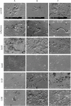
Osteoclastic differentiation and resorption is modulated by bioactive metal ions Co2+, Cu2+ and Cr3+ incorporated into calcium phosphate bone cements
- PMID: 28763481
- PMCID: PMC5538673
- DOI: 10.1371/journal.pone.0182109
Osteoclastic differentiation and resorption is modulated by bioactive metal ions Co2+, Cu2+ and Cr3+ incorporated into calcium phosphate bone cements
Abstract
Biologically active metal ions in low doses have the potential to accelerate bone defect healing. For successful remodelling the interaction of bone graft materials with both bone-forming osteoblasts and bone resorbing osteoclasts is crucial. In the present study brushite forming calcium phosphate cements (CPC) were doped with Co2+, Cu2+ and Cr3+ and the influence of these materials on osteoclast differentiation and activity was examined. Human osteoclasts were differentiated from human peripheral blood mononuclear cells (PBMC) both on the surface and in indirect contact to the materials on dentin discs. Release of calcium, phosphate and bioactive metal ions was determined using ICP-MS both in the presence and absence of the cells. While Co2+ and Cu2+ showed a burst release, Cr3+ was released steadily at very low concentrations (below 1 μM) and both calcium and phosphate release of the cements was considerably changed in the Cr3+ modified samples. Direct cultivation of PBMC/osteoclasts on Co2+ cements showed lower attached cell number compared to the reference but high activity of osteoclast specific enzymes tartrate resistant acid phosphatase (TRAP), carbonic anhydrase II (CAII) and cathepsin K (CTSK) and significantly increased gene expression of vitronectin receptor. Indirect cultivation with diluted Co2+ cement extracts revealed highest resorbed area compared to all other modifications and the reference. Cu2+ cements had cytotoxic effect on PBMC/osteoclasts during direct cultivation, while indirect cultivation with diluted extracts from Cu2+ cements did not provoke cytotoxic effects but a strictly inhibited resorption. Cr3+ doped cements did not show cytotoxic effects at all. Gene expression and enzyme activity of CTSK was significantly increased in direct culture. Indirect cultivation with Cr3+ doped cements revealed significantly higher resorbed area compared to the reference. In conclusion Cr3+ doped calcium phosphate cements are an innovative cement modification because of their high cytocompatibility and support of active resorption by osteoclasts.
Conflict of interest statement
Figures


References
-
- Bose S, Fielding G, Tarafder S, Bandyopadhyay A. Understanding of dopant-induced osteogenesis and angiogenesis in calcium phosphate ceramics. Trends Biotechnol. 2013;31: 594–605. doi: 10.1016/j.tibtech.2013.06.005 - DOI - PMC - PubMed
-
- Habibovic P, Barralet JE. Bioinorganics and biomaterials: bone repair. Acta Biomater. 2011;7: 3013–3026. doi: 10.1016/j.actbio.2011.03.027 - DOI - PubMed
-
- Mouriño V, Cattalini JP, Boccaccini AR. Metallic ions as therapeutic agents in tissue engineering scaffolds: an overview of their biological applications and strategies for new developments. J R Soc Interface R Soc. 2012;9: 401–419. doi: 10.1098/rsif.2011.0611 - DOI - PMC - PubMed
-
- Kulanthaivel S, Roy B, Agarwal T, Giri S, Pramanik K, Pal K, et al. Cobalt doped proangiogenic hydroxyapatite for bone tissue engineering application. Mater Sci Eng C Mater Biol Appl. 2016;58: 648–658. doi: 10.1016/j.msec.2015.08.052 - DOI - PubMed
-
- Birgani ZT, Gharraee N, Malhotra A, van Blitterswijk CA, Habibovic P. Combinatorial incorporation of fluoride and cobalt ions into calcium phosphates to stimulate osteogenesis and angiogenesis. Biomed Mater Bristol Engl. 2016;11: 015020 doi: 10.1088/1748-6041/11/1/015020 - DOI - PubMed
MeSH terms
Substances
LinkOut - more resources
Full Text Sources
Other Literature Sources

