. 2017 Aug 1;16(1):94.
doi: 10.1186/s12938-017-0390-3.

Video-tactile pneumatic sensor for soft tissue elastic modulus estimation

Affiliations

Video-tactile pneumatic sensor for soft tissue elastic modulus estimation

M M Gubenko et al. Biomed Eng Online. .

Abstract

Background: A new sensor for estimating elasticity of soft tissues such as a liver was developed for minimally invasive surgery application.

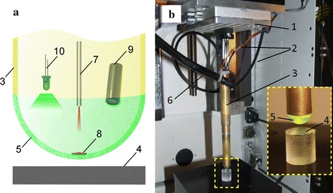
Methods: By measuring deformation and adjusting internal pressure of the pneumatic sensor head, the sensor can be used to do palpation (indentation) of tissues with wide range of stiffness. A video camera installed within the sensor shell is used to register the radius of the contact area. Based on finite element model simulations and the measured data, elastic modulus of the indented soft tissue can be calculated.

Results and conclusions: Three phantom materials, namely plastic, silicone and gelatin, with varied stiffness were tested. The experimental results demonstrated that the new sensor can obtain highly reliable data with error less than 5%. The new sensor might be served as an instrument in laparoscopic surgery for diagnosis of pathological tissues or internal organs.

Keywords: Indentation; Laparoscopy; Minimally invasive surgery; Tactile sensor; Young’s modulus.


Figures

Fig. 1
Fig. 1
a Schematic and b photograph of the sensor installed in a UMT, where 1 is the load cell, 2 commutation cords of the video camera and backlight, 3 cylindrical tube, 4 sample, 5 silicone shell of the sensor head, 6 compressed air supply, 7 optical proximity sensor, 8 mirror, 9 video camera, 10 LED
Fig. 2
Fig. 2
Scheme of the sensor shell (of radius R, mm) and the sample surface contact, where the dotted lines indicate un-deformed state at initial contact
Fig. 3
Fig. 3
Dependence of optical sensor readings U (volt) on displacement of shell central point under various air pressure conditions: 1 6 kPa, 2 15 kPa
Fig. 4
Fig. 4
Dependence of normal load P applied to the sensor on the shell displacement u s for materials with varying stiffness (gray markers 1, 3—a soft silicone sample, black markers 2, 4—a rigid sample) and varying air pressure: 1, 2—15 kPa; 3, 4—6 kPa
Fig. 5
Fig. 5
a The sensor indenting the glass installed on the reference camcorder. Images of the contact area from the built-in video camera (b) and from the reference camcorder (c). Black and white markers point the boundary of the contact area
Fig. 6
Fig. 6
a Contact scheme and a fragment of the finite elements mesh near the contact area. b Deformed shell in contact with gelatin phantom, stress distribution σz of the sample at h = 1.7 mm
Fig. 7
Fig. 7
Approximation of experimental data for gelatin by calculated curves. Family of curves with 15% step (E = 200 ± 30 kPa)
Fig. 8
Fig. 8
Distribution of contact pressure for gelatin phantom for various of h: 1.7 mm (curves 2, 4), 0.25 mm (curve 1), 0.15 mm (curve 3). p air = 15 kPa (curves 1, 2) and 6 kPa (curves 3, 4)
Fig. 9
Fig. 9
Dependence of the contact radius a on the displacement of the central point of the sensor shell u s (experimental points and calculated curves) for tests with gelatin (1) and silicone (2)

References

    1. Hatzinger M, Fesenko A, Buger L, Sohn M. Dimitrij Oscarovic Ott (1855–1929) “Ventroscopy”: his contribution to development of laparoscopy. Der Urologe Ausg A. 2013;52:1454–1458. doi: 10.1007/s00120-013-3224-3. - DOI - PubMed
    1. Manduca A, Oliphant TE, Dresner MA, Mahowald JL, Kruse SA, Amromin E, Felmlee JP, Greenleaf JF, Ehman RL. Magnetic resonance elastography: non-invasive mapping of tissue elasticity. Med Image Anal. 2001;5:237–254. doi: 10.1016/S1361-8415(00)00039-6. - DOI - PubMed
    1. Mariappan YK, Glaser KJ, Ehman RL. Magnetic resonance elastography: a review. Clin Anat. 2010;23:497–511. doi: 10.1002/ca.21006. - DOI - PMC - PubMed
    1. Evans A, Whelehan P, Thomson K, McLean D, Brauer K, Purdie C, Jordan L, Baker L, Thompson A. Quantitative shear wave ultrasound elastography: initial experience in solid breast masses. Breast Cancer Res. 2010;12:1–11. doi: 10.1186/bcr2787. - DOI - PMC - PubMed
    1. Shung KK. Diagnostic ultrasound: past, present, and future. J Med Biol Eng. 2011;31:371–374. doi: 10.5405/jmbe.871. - DOI

LinkOut - more resources