

Advances in microscale separations towards nanoproteomics applications
- PMID: 28765000
- PMCID: PMC6042839
- DOI: 10.1016/j.chroma.2017.07.055
Advances in microscale separations towards nanoproteomics applications
Abstract
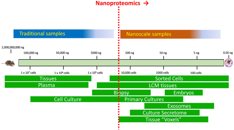
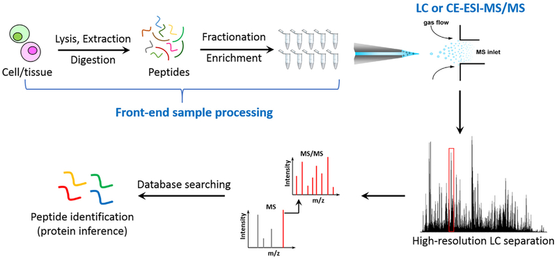
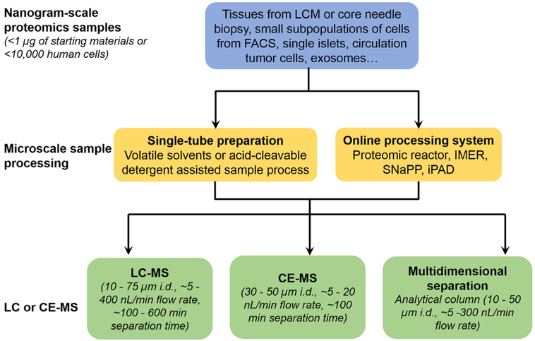
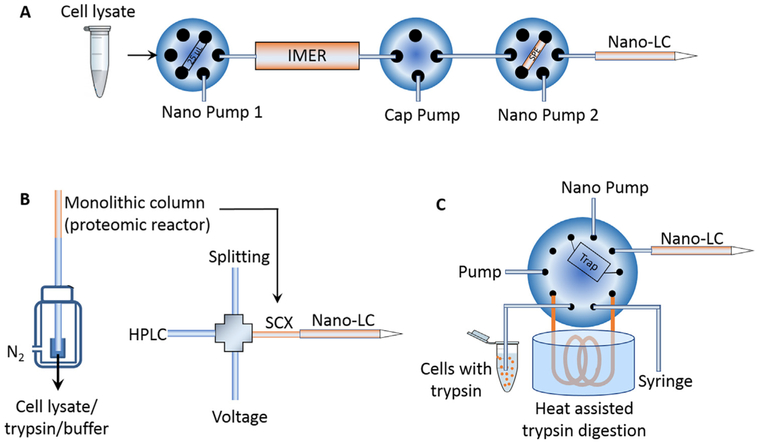
Microscale separation (e.g., liquid chromatography or capillary electrophoresis) coupled with mass spectrometry (MS) has become the primary tool for advanced proteomics, an indispensable technology for gaining understanding of complex biological processes. In recent decades significant advances have been achieved in MS-based proteomics. However, the current proteomics platforms still face an analytical challenge in overall sensitivity towards nanoproteomics applications for starting materials of less than 1μg total proteins (e.g., cellular heterogeneity in tissue pathologies). Herein, we review recent advances in microscale separation techniques and integrated sample processing strategies that improve the overall sensitivity and proteome coverage of the proteomics workflow, and their contributions towards nanoproteomics applications.
Keywords: Capillary electrophoresis; Mass spectrometry; Microscale separations; NanoLC; Nanoproteomics.
Copyright © 2017 Elsevier B.V. All rights reserved.
Conflict of interest statement
Conflict of interest
None.
Figures
References
-
- Mann M, Kulak NA, Nagaraj N, Cox J, The coming age of complete accurate, and ubiquitous proteomes, Mol. Cell 49 (2013) 583–590. - PubMed
-
- Aebersold R, Mann M, Mass-spectrometric exploration of proteome structure and function, Nature 537 (2016) 347–355. - PubMed
-
- Altelaar AF, Munoz J, Heck AJ, Next-generation proteomics: towards an integrative view of proteome dynamics, Nat. Rev. Genet 14 (2013) 35–48. - PubMed
-
- Larance M, Lamond AI, Multidimensional proteomics for cell biology, Nat. Rev. Mol. Cell Biol 16 (2015) 269–280. - PubMed
Publication types
MeSH terms
Substances
Grants and funding
LinkOut - more resources
Full Text Sources
Other Literature Sources
