Reversible central neural hyperexcitability: an electroencephalographic clue to hypocalcaemia
- PMID: 28765190
- PMCID: PMC5624021
- DOI: 10.1136/bcr-2017-220994
Reversible central neural hyperexcitability: an electroencephalographic clue to hypocalcaemia
Abstract
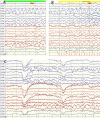
A 23-year-old male patient presented with cognitive decline and seizures. Examination revealed Chvostek's and Trousseau's signs. Investigations revealed hypocalcaemia, hyperphosphatemia and normal intact parathyroid hormone levels. Imaging showed calcifications in bilateral basal ganglia, thalamus and dentate nuclei. Interictal electroencephalogram showed theta range slowing of background activity and bilateral temporo-occipital, irregular, sharp and slow wave discharges, which accentuated during hyperventilation, photic stimulation and eye closure. Appearance of epileptiform discharges after eye closure, hyperventilation and photic stimulation may suggest presence of central neural hyperexcitability due to hypocalcaemia. These features may be an equivalent of peripheral neuromuscular hyperexcitability (Chvostek's and Trousseau's signs) that occurs in hypocalcaemia. The clinical and electroencephalographic features completely reversed with correction of serum calcium without antiepileptic medications. It is important for clinicians to recognise these reversible changes, as it can help to avoid misdiagnosis and long-term administration of antiepileptic becomes unnecessary.
Keywords: clinical neurophysiology; epilepsy and seizures.
© BMJ Publishing Group Ltd (unless otherwise stated in the text of the article) 2017. All rights reserved. No commercial use is permitted unless otherwise expressly granted.
Conflict of interest statement
Competing interests: None declared.
Figures


References
Publication types
MeSH terms
Substances
LinkOut - more resources
Full Text Sources
Other Literature Sources
Medical
