

Vascular Risk Factor Profiles Differ Between Magnetic Resonance Imaging-Defined Subtypes of Younger-Onset Lacunar Stroke
- PMID: 28765289
- PMCID: PMC5571884
- DOI: 10.1161/STROKEAHA.117.017813
Vascular Risk Factor Profiles Differ Between Magnetic Resonance Imaging-Defined Subtypes of Younger-Onset Lacunar Stroke
Abstract
Background and purpose: Differing associations of vascular risk factors with lacunar infarct have been reported, which is likely because of diagnostic differences and possible heterogeneity in the pathogenesis underlying lacunar infarction. In a large magnetic resonance imaging-verified cohort of lacunar infarct patients, we investigated the risk factor profile of lacunar infarction and magnetic resonance imaging characteristics.
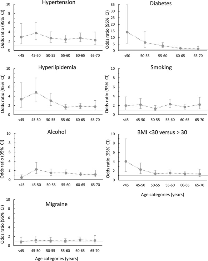
Methods: One thousand twenty-three patients with lacunar infarction (mean age, 56.7; SD, 8.5) were recruited from 72 stroke centers throughout the United Kingdom as part of the UK Young Lacunar Stroke DNA Study. Risk factor profiles were compared with 1961 stroke-free population controls with similar age. Furthermore, we tested risk factor profiles of lacunar stroke patients for association with the presence of multiple lacunar infarcts, white matter hyperintensities (WMH), and location of the acute lacunar infarct.
Results: Hypertension (odds ratio [OR], 2.21; 95% confidence interval [CI], 1.85-2.64), diabetes mellitus (OR, 2.10; 95% CI, 1.61-2.73), hyperlipidemia (OR, 1.74; 95% CI, 1.46-2.07), and smoking (OR, 1.65; 95% CI, 1.39-1.96) were independently associated in lacunar infarct patients compared with healthy controls. Patients with multiple lacunar infarcts were more likely to be men (OR, 2.53; 95% CI, 1.81-3.53) and have hypertension (OR, 1.54; 95% CI, 1.12-2.04) compared with patients with a single lacunar infarct, independent of other vascular risk factors. The presence of moderate-to-severe WMH versus no or mild WMH was independently associated with increased age (OR, 1.54; 95% CI, 1.12-2.04), hypertension (OR, 2.06; 95% CI, 1.44-2.95), and impaired renal function (OR, 0.90; 95% CI, 0.82-0.98).
Conclusions: In this magnetic resonance imaging-verified lacunar stroke population, we identified a distinct risk factor profile in the group as a whole. However, there were differing risk factor profiles according to the presence of multiple lacunar infarcts and confluent WMH. The association of hypertension, smoking, and renal impairment with the presence of multiple lacunar infarcts and confluent WMH might reflect a diffuse small vessel arteriopathy.
Keywords: population control; risk factors; smoking; stroke; white matter.
© 2017 The Authors.
Figures
References
-
- Román GC, Erkinjuntti T, Wallin A, Pantoni L, Chui HC. Subcortical ischaemic vascular dementia. Lancet Neurol. 2002;1:426–436. - PubMed
-
- Sudlow CL, Warlow CP. Comparable studies of the incidence of stroke and its pathological types: results from an international collaboration. International stroke incidence collaboration. Stroke. 1997;28:491–499. - PubMed
-
- Markus HS, Khan U, Birns J, Evans A, Kalra L, Rudd AG, et al. Differences in stroke subtypes between black and white patients with stroke: the south London ethnicity and stroke study. Circulation. 2007;116:2157–2164. doi: 10.1161/CIRCULATIONAHA.107.699785. - PubMed
-
- Arauz A, Murillo L, Cantú C, Barinagarrementeria F, Higuera J. Prospective study of single and multiple lacunar infarcts using magnetic resonance imaging: risk factors, recurrence, and outcome in 175 consecutive cases. Stroke. 2003;34:2453–2458. doi: 10.1161/01.STR.0000090351.41662.91. - PubMed
-
- Benavente OR, Pearce LA, Bazan C, Roldan AM, Catanese L, Bhat Livezey VM, et al. SPS3 Investigators. Clinical-MRI correlations in a multiethnic cohort with recent lacunar stroke: the SPS3 trial. Int J Stroke. 2014;9:1057–1064. doi: 10.1111/ijs.12282. - PubMed
Publication types
MeSH terms
Grants and funding
LinkOut - more resources
Full Text Sources
Other Literature Sources
Medical
