Tuberculosis presenting as broncho-oesophageal fistula in a young healthy man
- PMID: 28765480
- PMCID: PMC5623201
- DOI: 10.1136/bcr-2017-220821
Tuberculosis presenting as broncho-oesophageal fistula in a young healthy man
Abstract
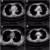
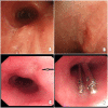
A 21-year-old Saudi man presented with a history of dysphagia and choking. CT scan of the chest showed clear evidence of chronic recurrent aspiration pneumonia in the left lung. It also showed a fistula connecting the left main bronchus to the oesophagus. Endoscopy showed clear opening on the oesophageal side. Bronchoscopy also confirmed the presence of a broncho-oesophageal fistula on the left bronchial side with the presence of secretions on swallowing. Bronchoalveolar lavage (BAL) was done and sent for mycobacterial tuberculosis culture. The fistula was closed with clips under endoscopic guidance, which alleviated his symptoms of dysphagia and choking. The BAL culture grew mycobacterial tubercle bacilli. The patient showed marked improvement after starting antitubercular therapy and was discharged to be followed up in the clinic.
Keywords: Gi-stents; Tb and other respiratory infections; endoscopy; infection (gastroenterology).
© BMJ Publishing Group Ltd (unless otherwise stated in the text of the article) 2017. All rights reserved. No commercial use is permitted unless otherwise expressly granted.
Conflict of interest statement
Competing interests: None declared.
Figures


References
-
- Diddee R, Shaw IH. Acquired tracheo-oesophageal fistula in adults. Continuing Education in Anaesthesia, Critical Care & Pain 2006;6:105–8. 10.1093/bjaceaccp/mkl019 - DOI
-
- Dieterich DT, Wilcox CM. Diagnosis and treatment of esophageal diseases associated with HIV infection. Practice Parameters Committee of the American College of Gastroenterology. Am J Gastroenterol 1996;91:2265–9. - PubMed
-
- Couraud L, Ballester MJ, Delaisement C. Acquired tracheoesophageal fistula and its management. Semin Thorac Cardiovasc Surg 1996;8:392–9. - PubMed
Publication types
MeSH terms
Substances
LinkOut - more resources
Full Text Sources
Other Literature Sources
Medical
