. 2017 Aug 1;7(1):6992.
doi: 10.1038/s41598-017-07117-8.

CDHR1 mutations in retinal dystrophies

Affiliations

CDHR1 mutations in retinal dystrophies

Katarina Stingl et al. Sci Rep. .

Abstract

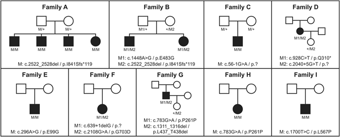
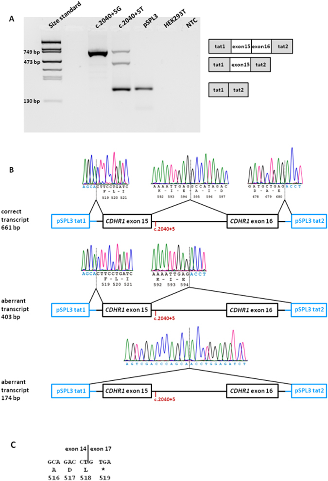
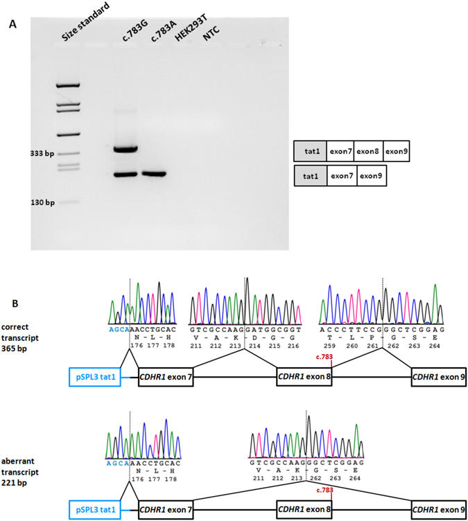
We report ophthalmic and genetic findings in patients with autosomal recessive retinitis pigmentosa (RP), cone-rod dystrophy (CRD) or cone dystrophy (CD) harboring potential pathogenic variants in the CDHR1 gene. Detailed ophthalmic examination was performed in seven sporadic and six familial subjects. Mutation screening was done using a customized next generation sequencing panel targeting 105 genes implicated in inherited retinal disorders. In one family, homozygosity mapping with subsequent candidate gene analysis was performed. Stringent filtering for rare and potentially disease causing variants following a model of autosomal recessive inheritance led to the identification of eleven different CDHR1 variants in nine index cases. All variants were novel at the time of their identification. In silico analyses confirmed their pathogenic potential. Minigene assays were performed for two non-canonical splice site variants and revealed missplicing for the mutant alleles. Mutations in CDHR1 are a rare cause of retinal dystrophy. Our study further expands the mutational spectrum of this gene and the associated clinical presentation.


Conflict of interest statement

The authors declare that they have no competing interests.

Figures

Figure 1
Figure 1
Pedigrees of all families. Disease-causing mutation(s) are given below each pedigree and the genotypes of all available family members.
Figure 2
Figure 2
In vitro splicing assay for the c.2040 + 5 G > T variant. (A) RT–PCR revealed three products in HEK 293 T cells transfected with the mutant construct and one product for the wild-type construct. Transfection with empty pSPL3 vector and untransfected cells served as controls. NTC, non template control. Schemes of the amplified products are presented on the right of the agarose gel. Grey boxes represent pSPL3 exons and white boxes CDHR1 exons. (B) Sequence analysis showing that one aberrant RT-PCR product from the mutant minigene construct results from skipping of exon 16 and the other aberrant RT-PCR product results from skipping of both exon 15 and 16. The position of the c.2040 + 5 G > T variant is depicted in red. (C) Consequence of the skipping of exon 15 and 16 on protein level. Mutant CDHR1 transcript lacking exons 15 and 16 will contain a premature stop codon (protein translation is shown below DNA sequence in one letter amino-acid code).
Figure 3
Figure 3
In vitro splicing assay for the c.783 G > A variant. (A) RT-PCR revealed two products in HEK 293 T cells transfected with the wild-type construct and a single RT-PCR product for the mutant construct. Untransfected cells served as control. NTC, non-template control. Schemes of the amplified products are presented on the right of the agarose gel. Grey boxes represent pSPL3 exons and white boxes CDHR1 exons. (B) Sequence analysis showing that the aberrant product results from skipping of exon 8. The position of the c.783 variant is depicted in red. The junction of CDHR1 exon 9 and pSPL3 tat2 could not be shown since a reverse PCR primer was used that binds within exon 9.
Figure 4
Figure 4
Summary of clinical findings. The composite image shows the most important available clinical findings. Each row represents a single patient with the following information in each column: Family (affiliation to the family according to Fig. 1); clinical diagnosis (CRD, CD or RP); genotype; age of onset (as reported subjectively by the patient); age (at examination); first symptom (first manifestation of the disease as reported by the patient); BCVA (best corrected visual acuity); night blindness, photophobia, color vision defect (“Y” for present, “N” for absent); fullfield ERG; visual field (if available); fundus image (if available); fundus autofluorescence image (if available).

References

    1. Thiadens AA, et al. Clinical course, genetic etiology, and visual outcome in cone and cone-rod dystrophy. Ophthalmology. 2012;119:819–826. doi: 10.1016/j.ophtha.2011.10.011. - DOI - PubMed
    1. Michaelides M, Hardcastle AJ, Hunt DM, Moore AT. Progressive cone and cone-rod dystrophies: phenotypes and underlying molecular genetic basis. Surv Ophthalmol. 2006;51:232–258. doi: 10.1016/j.survophthal.2006.02.007. - DOI - PubMed
    1. Weleber, R. G. Retinitis pigmentosa and allied disorders in Retina (ed. Ryan, S. J.) 335–466 (Mosby, 1994).
    1. Nollet F, Kools P, van Roy F. Phylogenetic analysis of the cadherin superfamily allows identification of six major subfamilies besides several solitary members. J Mol Biol. 2000;299:551–572. doi: 10.1006/jmbi.2000.3777. - DOI - PubMed
    1. Ahmed ZM, et al. Mutations of the protocadherin gene PCDH15 cause Usher syndrome type 1F. Am J Hum Gen. 2001;69:25–34. doi: 10.1086/321277. - DOI - PMC - PubMed

Publication types

MeSH terms

LinkOut - more resources