. 2017 Aug 1;7(1):6976.
doi: 10.1038/s41598-017-07072-4.

Calcium Ion Induced Structural Changes Promote Dimerization of Secretagogin, Which Is Required for Its Insulin Secretory Function

Affiliations

Calcium Ion Induced Structural Changes Promote Dimerization of Secretagogin, Which Is Required for Its Insulin Secretory Function

Jae-Jin Lee et al. Sci Rep. .

Abstract

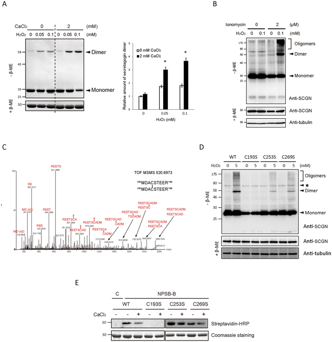
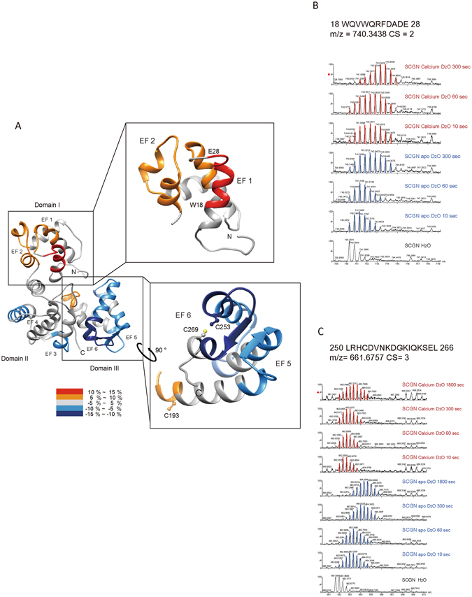
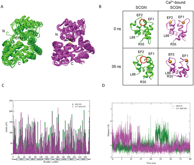
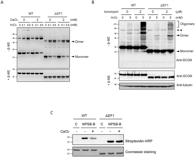
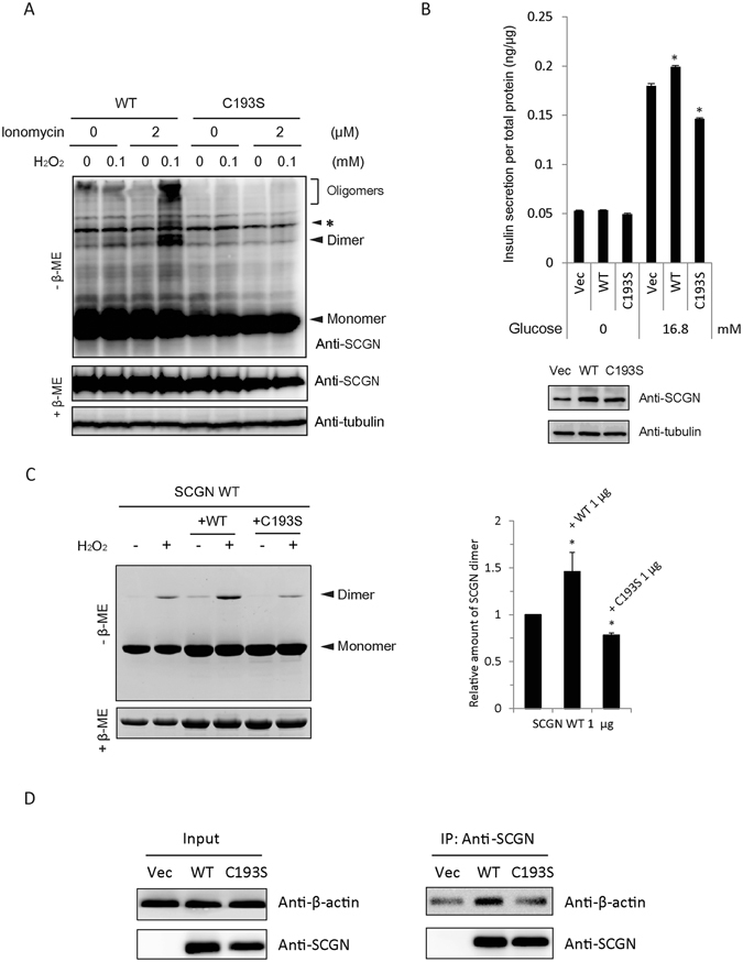
Secretagogin (SCGN), a hexa EF-hand calcium binding protein, plays key roles in insulin secretion in pancreatic β-cells. It is not yet understood how the binding of Ca2+ to human SCGN (hSCGN) promotes secretion. Here we have addressed this question, using mass spectrometry combined with a disulfide searching algorithm DBond. We found that the binding of Ca2+ to hSCGN promotes the dimerization of hSCGN via the formation of a Cys193-Cys193 disulfide bond. Hydrogen/deuterium exchange mass spectrometry (HDX-MS) and molecular dynamics studies revealed that Ca2+ binding to the EF-hands of hSCGN induces significant structural changes that affect the solvent exposure of N-terminal region, and hence the redox sensitivity of the Cys193 residue. These redox sensitivity changes were confirmed using biotinylated methyl-3-nitro-4-(piperidin-1-ylsulfonyl) benzoate (NPSB-B), a chemical probe that specifically labels reactive cysteine sulfhydryls. Furthermore, we found that wild type hSCGN overexpression promotes insulin secretion in pancreatic β cells, while C193S-hSCGN inhibits it. These findings suggest that insulin secretion in pancreatic cells is regulated by Ca2+ and ROS signaling through Ca2+-induced structural changes promoting dimerization of hSCGN.


Conflict of interest statement

The authors declare that they have no competing interests.

Figures

Figure 1
Figure 1
Calcium binding of hSCGN promotes H2O2 induced dimerization via C193-C193 disulfide linkage. (A) Recombinant hSCGN was incubated with 0 or 2 mM CaCl2 at R.T. for 15 min followed by H2O2 treatment in indicated concentration at 37°C for 1 h. Proteins were detected with coomassie blue-staining (left panel) and dimer formations were quantified and normalized with reduced form of SCGN (right panel). Data are presented as mean ± SD of three experiments (*P < 0.05, Student’s t-test). (B) NIT-1 insulinoma cells overexpressing hSCGN WT were pre-incubated with 0 or 2 μM ionomycin for 5 min followed by incubation with 0 or 100 μM H2O2 for 30 min at 37 °C. Total protein lysates were analyzed by Western blots using anti-SCGN or anti-α-tubulin antibody. (C) Tandem mass spectrum of C193-C193 disulfide linked peptide in band #7 and 8 of Supplementary Fig. S2A. (D) HeLa cells overexpressing SCGN WT, and Cys mutant (C193S, C253S, C269S) were treated with 0, 5 mM H2O2 for 10 min at 37°C. The total lysates were separated on non-reducing and reducing SDS PAGE and hSCGN and tubulin were detected by Western analysis. Tubulin was used for loading control. Non-specific bands were indicated as *. (E) hSCGN WT, and Cys mutants were incubated with 2 mM CaCl2 at R.T. for 15 min followed by treatment with 1 mM NPSB-B at R.T. for 2 h. Proteins were separated on reducing SDS-PAGE and detected by streptavidin-HRP. Coomassie staining gel showing amount of gel loaded proteins. Full-length gels and blots are in Supplementary Fig. S6.
Figure 2
Figure 2
Structural changes in apo and Ca2+-bound hSCGN analyzed by hydrogen/deuterium exchange mass spectrometry. (A) Overlay of differential HDX data onto the structures of human SCGN structure which were modeled in silico with the MODELLER 9.9 program. Percentage difference in HDX between apo and Ca2+ bound hSCGN is colored according to the key. The differential deuterium exchange patterns of hSCGN peptide sequences representing EF-hand and Cys residue in N-terminal (B) and C-terminal (C) peptides of hSCGN.
Figure 3
Figure 3
Dynamics of apo and six Ca2+-bound hSCGN in molecular dynamics simulation. (A) The coordinates of apo-hSCGN and Ca2+-bound hSCGN saved every 5 ns during MD simulations are superimposed, respectively. (B) Snapshots of the domain I of apo-hSCGN and Ca2+-bound hSCGN at 0 ns and 35 ns. Ca2+ ions are drawn as orange spheres. The conformation of the red circled area is a β-sheet at 0 ns when analyzed by STRIDE (http://webclu.bio.wzw.tum.de/cgi-bin/stride/stridecgi.py). (C) The average SASA values for every residue of 400 structures saved every 100 ps during 40 ns simulation. (D) The distances between Cys253 and Cys269 during simulation for apo-hSCGN (green) and Ca2+-bound hSCGN (magenta).
Figure 4
Figure 4
N-terminus of hSCGN is essential for calcium response. (A) Purified hSCGN WT and ΔEF1 protein were pre-treated with 0, 2 mM CaCl2 for 15 min at R.T. and then incubated with the indicated concentrations of H2O2 for 1 h at 37°C. Coomassie blue-stained SDS-PAGE gels under non-reducing (−β-ME) and reducing (+β-ME) condition. (B) HeLa cells overexpressing hSCGN WT and ΔEF1 were treated with 0 or 2 μM ionomycin for 5 min and with 0 or 5 mM H2O2 for 30 min at 37 °C. Cell lysates were subjected to Western analysis using anti-hSCGN or anti-α-tubulin antibody. Non-specific bands were indicated as *. (C) Recombinant hSCGN WT and ΔEF1 proteins were incubated with 0 or 2 mM CaCl2 for 15 min, and with 1 mM NPSB-B for 2 h at R.T. Labeled samples in gel sample buffer containing 10 mM NEM were detected with streptavidin-HRP. Full-length gels and blots are in Supplementary Fig. S7.
Figure 5
Figure 5
Dimerization of hSCGN is required for its role in insulin secretion. (A) NIT-1 insulinoma cells overexpressing hSCGN WT and C193S mutant were pre-incubated with 0 or 2 μM ionomycin for 5 min followed by incubation with 0 or 100 μM H2O2 for 30 min at 37 °C. Proteins were separated under non-reducing and reducing conditions on SDS PAGE and detected with Western analysis using anti-SCGN antibody. Non-specific bands were indicated as *. (B) NIT-1 cells overexpressing hSCGN WT or C193S mutant were starved with glucose-free HBSS for 2 h followed by stimulation with 16.8 mM glucose for 25 min. The amount of secreted insulin was measured and normalized to total cell protein concentration. Whole cell lysates were subjected to Western analysis with anti-SCGN or anti-α-tubulin antibody. (C) Dimer formations of hSCGN WT, and hSCGN WT mixed with same amount of hSCGN WT or C193S proteins were measured after treating with 2 mM CaCl2 for 15 min at R.T. followed by incubation with 0 or 1 mM H2O2 for 1 h at 37°C. Proteins were detected with coomassie blue-staining (left panel) and were quantified and normalized with reduced form of SCGN (right panel). (D) hSCGN C193S fails to interact with cytoskeletal actin in MDA-MB-231 cells. MDA-MB-231 cells overexpressing SCGN WT and C193S mutant were lysed with a lysis buffer containing protease inhibitors. Cell lysates were immunoprecipitated with anti-SCGN antibody and the immune complexes were separated on SDS-PAGE and detected with western analysis using anti-actin and anti-SCGN antibodies. Data information: In (B,C), data are expressed as the mean ± SD of three experiments (*P < 0.05, Student’s t-test). Full-length gels and blots are presented in Supplementary Fig. S8.

References

    1. Wagner L, et al. Cloning and expression of secretagogin, a novel neuroendocrine- and pancreatic islet of Langerhans-specific Ca2+-binding protein. The Journal of biological chemistry. 2000;275(32):24740–24751. doi: 10.1074/jbc.M001974200. - DOI - PubMed
    1. Romanov RA, et al. A secretagogin locus of the mammalian hypothalamus controls stress hormone release. EMBO J. 2015;34(1):36–54. doi: 10.15252/embj.201488977. - DOI - PMC - PubMed
    1. Mulder J, et al. Secretagogin is a Ca2+-binding protein specifying subpopulations of telencephalic neurons. Proc Natl Acad Sci USA. 2009;106(52):22492–22497. doi: 10.1073/pnas.0912484106. - DOI - PMC - PubMed
    1. Attems J, et al. Clusters of secretagogin-expressing neurons in the aged human olfactory tract lack terminal differentiation. Proc Natl Acad Sci USA. 2012;109(16):6259–6264. doi: 10.1073/pnas.1203843109. - DOI - PMC - PubMed
    1. Bauer M, et al. Protein networks involved in vesicle fusion, transport, and storage revealed by array-based proteomics. Methods Mol Biol. 2011;781:47–58. doi: 10.1007/978-1-61779-276-2_3. - DOI - PubMed

Publication types